Flatfoot (pes planus) is an overpronation of the foot, resulting in a lower or absent arch. Patients with flatfoot fall into two groups: adult and pediatric (child). While both groups exhibit flatfoot, the causes and treatments may differ.

Diagnosing and Treating Adult Flatfoot
Adults may develop flatfoot due to failure of the posterior tibial tendon. This is a large tendon originating in the leg and attaching to the navicular bone in the foot. This tendon helps support the arch and helps propel a person forward when they walk. Over time, the tendon can stretch and even tear, allowing the arch to fall.
With flatfoot, simple actions – standing on your tiptoes, walking, traveling up and downstairs, or standing – can start to become painful. Adult flatfoot is also accompanied by swelling and weakness when trying to turn the ankle inward.
X-rays can display change in the foot structure such as a fallen arch, but they often show no change at all. Patients with long-standing posterior tendinitis may show signs of arthritis. MRI can help determine if tendinitis is present and the severity of the condition.
90 percent of patients with flatfoot can be successfully treated with non-steroidal anti-inflammatory drugs (NSAIDs), changes in shoe gear, arch supports, and in some cases, physical therapy. Patients experiencing severe pain may need a walking (CAM) boot.
For patients showing no improvement with any of the above treatments, surgical correction may be an option. The goal of flatfoot surgery is to restore the strength and integrity of the tendon and improve the structure of the arch.
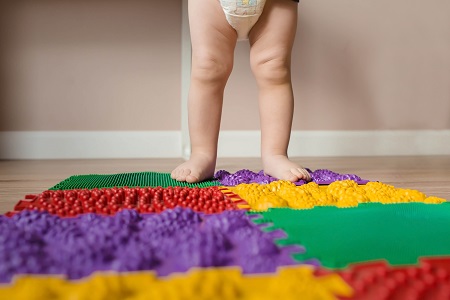
Diagnosing and Treating Pediatric Flatfoot
Most children are born with flatfoot and the arch begins to develop over time; however, some children never develop an arch. Pediatric flatfoot may develop due to hypermobility (double-jointedness). A lump may also develop inside the foot, indicating an accessory navicular bone.
Although many pediatric patients are asymptomatic, some may experience foot, ankle, and leg pain. Pain is particularly noticeable during running and playing.
X-rays can help determine the severity, the type of flatfoot, and the presence of an accessory navicular bone.
Asymptomatic children benefit from over-the-counter support, while symptomatic children benefit from a custom orthotic. Children who have activity restrictions due to severe pain and have failed conservative treatment may benefit from surgical correction. Surgical correction includes bone grafting, lengthening a tight heel Achilles tendon, and removing an accessory navicular.
more recommended stories
 Hypertensive Disorders of Pregnancy: Role of Daily Activity
Key Points Summary Limiting sedentary time.
 Climate Change Drives Dengue Outbreaks Globally
Key Takeaways Extreme weather significantly increases.
 PFK Enzyme Dual Role in Metabolism and Cell Cycle
Key Highlights Phosphofructokinase (PFK Enzyme) shows.
 Food Tolerance Mechanism: How T Cells Prevent Allergies
Key Summary Researchers at Stanford University.
 Endometriosis Screening Tool May Cut Diagnosis Delays
Key Points Researchers from the University.
 Diabetic Kidney Disease: Combo Therapy Targets Zombie Cells
Key Points Researchers from Mayo Clinic.
 Dance for Healthy Aging: Study Highlights Benefits
Key Points at a Glance A.
 Breast Cancer Prognosis Linked to High-Fat Diet
Key Points A high-fat diet accelerated.
 Advanced Prostate Cancer and Serial ctDNA Analysis
Key Takeaways Serial liquid biopsies using.
 Tuberculosis Breakthrough with Experimental Antibiotics
Key Takeaways Experimental antibiotics disrupt a.
Leave a Comment