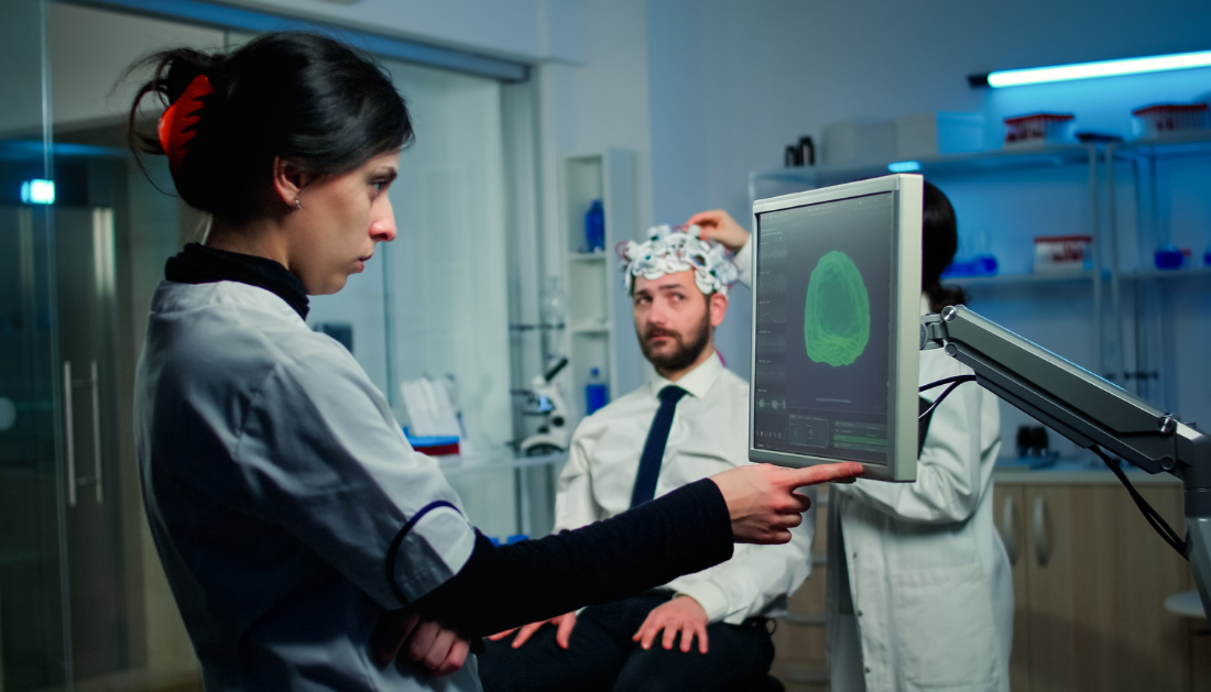

A wearable ultrasonic patch created by engineers at the University of California, San Diego has the potential to provide continuous, non-invasive monitoring of cerebral blood flow. For the first time in wearable technology, the soft, flexible patch may be worn comfortably on the temple to offer three-dimensional data on cerebral blood flow.
The researchers published their novel technology in Nature under the direction of Sheng Xu, a professor in the UC San Diego Jacobs School of Engineering’s Aiiso Yufeng Li Family Department of Chemical and Nano Engineering.
Transcranial Doppler ultrasonography, the current clinical standard, is much inferior to the wearable ultrasound patch. An ultrasound probe must be held against the patient’s head by a qualified technician in order to use this approach. There are drawbacks to the procedure, though. Because it is operator-dependent, the operator’s skill level will determine how accurate the measurement is. Moreover, it is not feasible for continuous usage.
Xu’s group created a tool that gets around these obstacles. Throughout a patient’s hospital stay, their wearable ultrasonography patch provides a hands-free, reliable, and comfortable solution that can be worn continually.
In Xu’s lab, Sai Zhou, a Ph.D. candidate in materials science and engineering, is co-first author of the work. “The continuous monitoring capability of the patch addresses a critical gap in current clinical practices,” Zhou said. Cerebral blood flow is typically measured at specified periods of the day, and such readings may not accurately reflect what occurs during the remainder of the day. Unnoticed variations may occur in between measurements. This gadget may provide information that is essential for prompt intervention if a patient is about to suffer a stroke in the middle of the night.”
According to Geonho Park, a Ph.D. student in Xu’s lab studying chemical and nanoengineering and another co-first author of this work, patients undergoing and recuperating from brain surgery can also benefit from this technique.
The patch is made of silicone elastomer and has multiple layers of flexible electronics embedded in it. It is about the size of a postage stamp. One layer is made up of a collection of tiny piezoelectric transducers that, when electrically activated, generate ultrasonic waves and receive waves that are reflected from the brain. A copper mesh layer consisting of spring-shaped wires is another essential element that improves signal quality by reducing interference from the wearer’s body and surroundings. Elastic electrodes make up the remaining layers.
The patch is wired to a computer and power source in order to function. The researchers included ultrafast ultrasound imaging into the device in order to accomplish 3D monitoring. Ultrafast imaging records thousands of images per second, in contrast to normal ultrasound’s 30 photos per second. Because of the heavy reflection of the skull, the piezoelectric transducers in the patch would ordinarily have poor signal intensity, making this fast frame rate essential for obtaining reliable data.
After that, the data are post-processed using unique algorithms to recreate 3D details like the major arteries of the brain’s size, angle, and location.
“The cerebral vasculature is a multibranched, intricate system of vessels. A second co-first author of this work and a Ph.D. candidate in materials science and engineering at Xu’s group, Xinyi Yang, stated, “To get the full picture and obtain more accurate measurements, you need a device capable of capturing this three-dimensional information.”
The ability of the patch to assess blood flow velocities—peak systolic, mean flow, and end diastolic velocities—in the primary arteries supplying the brain was examined in this study on 36 healthy participants. Participants performed blood-flow-affecting exercises like grasping with their hands, holding their breath, and reading. The measures taken with the patch and a traditional ultrasonography probe agreed quite well.
The patch will then be tested on individuals with neurological disorders that affect cerebral blood flow by the researchers in partnership with medical professionals at UC San Diego School of Medicine. Xu is a co-founder of Softsonics, a startup firm aimed at commercializing this technology.
For more information: Transcranial volumetric imaging using a conformal ultrasound patch, Nature, https://dx.doi.org/10.1038/s41586-024-07381-5
more recommended stories
 High-Fat Diets Cause Damage to Metabolic Health
Key Points Takeaways High-fat and ketogenic.
 Can Too Many Antioxidants Harm Future Offspring?
Key Takeaways High-dose antioxidant supplementation in.
 Human Antibody Drug Response Prediction Gets an Upgrade
Key Takeaways A new humanized antibody.
 Dietary Melatonin Linked to Depression Risk: New Study
Key Summary Cross-sectional analysis of 8,320.
 Type 2 Diabetes Risk Identified by Blood Metabolites
Key Takeaways (Quick Summary) Researchers identified.
 Microglia Neuroinflammation in Binge Drinking
Key Takeaways (Quick Summary for HCPs).
 Durvalumab in Small Cell Lung Cancer: Survival vs Cost
Key Points at a Glance Durvalumab.
 Rising Chagas Parasite Detected in Borderland Kissing Bugs
Key Takeaways (At a Glance) Infection.
 Can Ketogenic Diets Help PCOS? Meta-Analysis Insights
Key Takeaways (Quick Summary) A Clinical.
 Ancient HHV-6 Genomes Confirm Iron Age Viral Integration
Key Takeaways for HCPs Scientists reconstructed.

Leave a Comment