Study: Cutting-edge portable MRI technology set for human trials
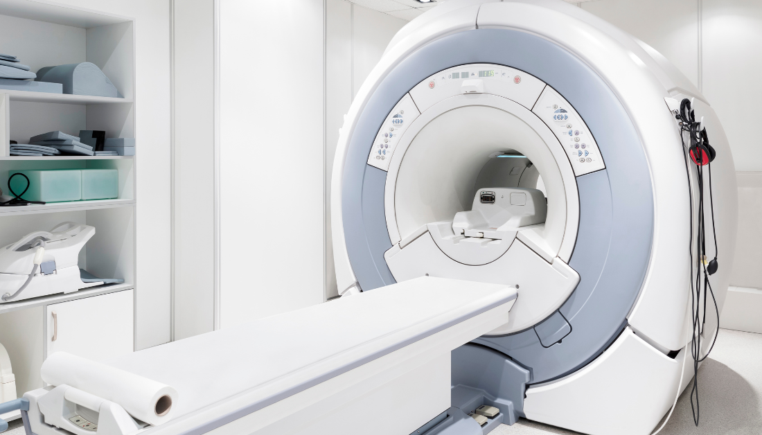
A team led by University of Minnesota academics has invented a Portable MRI technology, new type of Magnetic Resonance Imaging (MRI) scanner that provides images of comparable quality to traditional machines but taking up significantly less space.
This innovative machine disassembles into four pieces and fits onto the bed of a truck, allowing it to be transpmriorted to distant villages where residents may not have access to MRI scanners. A typical MRI machine weighs 4,500 kg; the new one weighs only 400.
Michael Garwood, associate director of the Center for Magnetic Resonance Research (CMRR) and lead researcher on the project, said 90% of the world’s population does not have access to an MRI machine.
Dependable access to an MRI machine is important for helping diagnose, monitor and treat injuries and diseases, according to Garwood.
“This is the motivating factor for all this work, there’s just not enough MRIs,” Garwood said. “You can’t access 90% of the world’s population with it and it’s time to do something about that.”
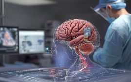
Garwood’s portable MRI, unlike traditional machines, only scans the head, which is one of the reasons this new machine can be so compact while yet functioning effectively.
The machine resembles a salon-style hooded hair dryer. The patient sits in a chair that has been lifted so that their head is completely beneath the dome. During the treatment, a strip of glass is inserted at eye level for the patient to look out of, minimizing symptoms of claustrophobia.
Garwood said keeping children in mind when designing the machine was a priority. Kids can get scared when going through the larger machine, so Garwood wanted to make the MRI process more inviting and less intimidating.
Garwood said he originally designed the machine to run at 1.5 tesla, a unit of measurement for magnetic flux, just like a standard MRI machine. However, by using paraffin wax as a binding agent instead of epoxy, the machine was able to run at about half power.
Even though it is a lower operating field than standard MRI machines, Garwood said, his machine will not affect the images it generates and is safe for human use.
Garwood demonstrated the machine’s inner workings during a personal tour of the lab. When asked for a photo, he chose to raise himself into the machine while the magnet was turned on, essentially completing the first unofficial human testing.
Despite the fact that Garwood did not scan his brain while having his photo taken, it was a display of how close the team is to starting actual human testing. Garwood anticipates that controlled human studies will commence within the next two months.
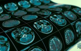
Initially, the researchers used the gadget to scan water bottles and lemons. These scans produced high-quality images without the requirement for artificial intelligence (AI).
Researchers from the University, Yale, and Columbia, as well as scientists from Brazil and New Zealand, were needed to collaborate on the study.
Alberto Tannus, one of Garwood’s Brazilian professor colleagues, designed a spectrometer that serves as the machine’s brain. It was created independently of Garwood’s initiat
Tannus said the research was hard to conduct because of funding. In Brazil specifically, Tannus said he was dealing with a shy budget.
“Something that I learned is if you can do it with this very low budget here in Brazil, you can do it with any amount of money anywhere in the world,” Tannus said.
Ben Parkinson, a researcher on Garwood’s team from New Zealand, said when the team had to design a magnet for the machine, they had to use a different process because of how small their machine is compared to a traditional MRI.
Garwood’s machine can produce clinical-quality images at a smaller size because it only gathers images from the head. Its smaller size and lower tesla also allow it to have a less uniform magnetic field than a standard MRI machine.
Parkinson said another difference between their portable MRI machine and a standard MRI is the temperature control. A standard machine needs the coils inside the magnet to be cooled off in a bath of liquid helium.
Garwood’s Portable MRI uses a high-temperature superconducting (HTS) magnet, which helps the coils inside cool faster and more efficiently.
Cristoph Juchem, an associate professor at Columbia University in New York, designed the hardware for the coils inside of the magnet.
A traditional MRI machine uses gradient coils to predictably manipulate the magnetic fields and typically relies on a set of three coils. The array Juchem developed houses 31 coils that can be controlled individually to create more flexible and dynamic patterns.
Juchem said getting a project like this off the ground is a hard enough task and is very excited they are nearing human trials.
“The fact that we get clean images, even though it’s not human yet, means we’re certainly on the right track and now the next goal is to do this on people like you and me,” Juchem said.
ive and was intended to be used as a research tool.
The spectrometer, which is housed on a 1-inch-by-1-inch processor chip, connects logical ports. The spectrometer assists the machine in getting information where it needs to go, similar like phoning a general customer service number and being re-routed to a more particular department.
 Phage Therapy Study Reveals RNA-Based Infection Control
Key Takeaways (Quick Summary) Researchers uncovered.
 Safer Allogeneic Stem Cell Transplants with Treg Therapy
A new preclinical study from the.
 AI in Emergency Medicine and Clinician Decision Accuracy
Emergency teams rely on rapid, accurate.
 Innovative AI Boosts Epilepsy Seizure Prediction by 44%
Transforming Seizure Prediction in Epilepsy Seizure.
 Hypnosis Boosts NIV Tolerance in Respiratory Failure
A New Approach: Hypnosis Improves NIV.
 Bee-Sting Microneedle Patch for Painless Drug Delivery
Microneedle Patch: A Pain-Free Alternative for.
 AI Reshapes Anticoagulation in Atrial Fibrillation Care
Understanding the Challenge of Atrial Fibrillation.
 Hemoglobin as Brain Antioxidant in Neurodegenerative Disease
Uncovering the Brain’s Own Defense Against.
 Global Data Resource for Progressive MS Research (Multiple Sclerosis)
The International Progressive MS Alliance has.
 AI Diabetes Risk Detection: Early T2D Prediction
A new frontier in early diabetes.
Leave a Comment