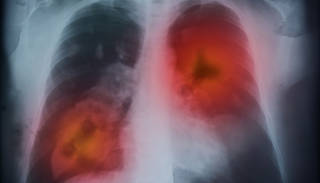

Lung cancer is a known killer. According to the American Cancer Society, the disease kills more than 125,000 Americans each year, more than colon, breast, and prostate cancer combined. For many years, the outlook was grim, and survival rates were depressingly low.
However, clinicians and scientists at the OHSU Knight Cancer Institute say this does not have to be the case. Lung cancer is treatable and curable if found early.
Teams at the OHSU Knight Cancer Institute are initiating a project to transform the way lung cancer is treated using cutting-edge technologies. The effort has the potential to save thousands of lives and significantly reduce the disease’s burden in Oregon.
The project, which will be launched on November 11, National Lung Cancer Screening Day, comprises four components:
Detecting cancer earlier through annual screening
A high-tech robot that looks deeper inside the lung, where most cancers begin.
More patient assistance, such as nurse navigators
More methods to help people quit smoking — anyone can acquire lung cancer, but individuals who smoke or have smoked are at a higher risk.
Because of advancements like these, lung cancer is more likely to be detected at an early stage, increasing patients’ five-year survival rate to more than 90%. This is in stark contrast to the outcomes when cancer is detected late, when survival rates are below 10%.
“Annual low-dose chest CT scanning is a fast, easy and painless screening test for lung cancer,” says radiologist Chara Rydzak, M.D., Ph.D., one of three co-directors of the new program and an associate professor diagnostic radiology, OHSU School of Medicine. “It allows us to diagnose lung cancers quickly and at the earliest stages possible. This improves survival and cure rates for patients, giving them more time to enjoy life and spend time with their loved ones.”
Katie Putnam, M.D., M.P.H., resident physician in family medicine, and Peter Lee, M.D., M.H.S., assistant professor of medicine (pulmonary and critical care medicine), OHSU School of Medicine, are the program’s other directors.
“We can make a huge difference if we catch this early,” says Ann Spencer, RN, a nurse navigator. “Thousands of people in Oregon could benefit from screening.”
Screening helps save lives
Lung cancer has no signs in its early stages – there is no lump to feel or mole to notice. As a result, it is frequently not identified until it has progressed to an advanced stage and has spread throughout the body. According to the National Cancer Institute, a part of the National Institutes of Health, just 25% of all cases are detected at stage I.
Low-dose CT scans, on the other hand, can detect lung cancer long before patients perceive any symptoms. Patients had to pay for their own scans until 2015, when Medicare began to pay for screening; presently, most insurance plans include screening for high-risk individuals.
The United States Preventive Services Task Force announced new guidelines in 2021 on who would benefit the most: anyone aged 50 to 80 with a smoking history of 20 pack-years (a pack a day for 20 years) who is still smoking or has quit within the last 15 years. The revised suggestions increased the number of Black and female screening candidates.
However, adoption has been gradual. In 2022, only 7% of the eligible population in Oregon was examined. Researchers have identified two major barriers: access and “therapeutic nihilism,” or the false idea that lung cancer is incurable.
To overcome these obstacles, OHSU has undertaken an ambitious program to screen more people.
“Smokers and ex-smokers should absolutely get screened,” says Eliza Kaiser, RN, BSN, CCRN, a nurse navigator. “Early detection is a game-changer.”
Nurse navigators, such as Kaiser and Spencer, assist patients in registering for screening, scheduling appointments, quitting smoking, and coordinating their treatment among the various specialists they encounter. “We’re here for you every step of the way,” says Kaiser.
A low-dose CT scan usually takes 15 minutes. If a scan reveals something questionable, nurse navigators can assist patients in taking the next step: determining whether or not they have cancer.
A GPS system for the lungs
The passageway splits deep inside the maze, in a gleaming tunnel speckled with burgundy reds and rosy pinks. Lee, program co-director and interventional pulmonologist, checks his coordinates above the pleasant whirling of fans and the chirp of biomonitors. He’ll go with the left fork. He expertly navigates forward using the trackball and scroll wheel. It looks like a scenario from Star Wars, but Lee and his crew are in an operating room at OHSU, using a robot to guide a super-thin scope through a patient’s lung.
A lung nodule is a spot on the lung that can be seen on a scan. The majority of nodules are insignificant; small ones are typically the product of scar tissue from an old infection and are not cause for alarm. A larger nodule is more likely to be cancer and, if detected early, can be treated surgically. But first, the team must determine whether it is cancer or not. This necessitates a bronchoscopy, in which Lee and his team use the Ion robotic-assisted navigation device, affectionately dubbed “Rosey,” to reach deep into the lung and collect samples from suspicious nodules.
Bronchoscopy is a difficult operation because the airway is a complex labyrinth of forking channels that narrow and get more convoluted as they branch deeper and deeper inside the lung. Rydzak, a thoracic radiologist, reviews a CT scan and pinpoints the exact position of the nodule to plot a path through the maze. Rosey employs cutting-edge technology to generate a precise, three-dimensional map of the best route – sort of like GPS for the lung.
“It’s a powerful tool,” adds Lee. “The Ion is far more accurate and safe than older navigation systems.” We can reach nodules in areas of the lung where other equipment cannot.”
Lee delicately slides the scope through the twists and turns of the airway after the patient has been securely anesthetized. Rosey employs optical-fiber technology to keep the scope steady and on track. When the scope reaches the nodule, Lee uses a cryogenic tool to freeze and retrieve a small sample for the pathology team to examine. If cancer is discovered, the team can do additional testing to determine which sort of treatment will be most beneficial. In some circumstances, they can adapt the medication to the cancer’s precise genomic profile.
breathing space
Surgery used to entail removing an entire lung by a major incision in the early days of lung cancer treatment. OHSU thoracic surgeons have been removing early-stage lung tumors with very small incisions and minimally invasive procedures for many years, preserving more of the patient’s lung. These life-saving procedures can benefit more individuals if they are detected earlier.
We know how to do big, complicated lung surgeries for advanced lung cancer, and when it is needed, we are here,” says Paul Schipper, M.D., FACS, FACCP, professor of surgery (cardiothoracic surgery) at OHSU. “With early detection, however, we finally get the chance to cure lung cancer in a lot more people and spare more healthy lung tissue so they can get back to their lives.”
“Identifying lung cancers earlier allows us, as surgeons, to surgically treat the cancer effectively while preserving lung function for our patients,” says Ruchi Thanawala, M.D., M.S., FACS, assistant professor of cardiothoracic surgery at OHSU.
Early detection of lung cancer also allows for more potent types of radiation therapy.
Over the past two decades, advances in imaging, radiation delivery, and computer modeling have significantly advanced radiotherapy for lung cancer,” says Josh Walker, M.D., Ph.D., assistant professor of cell, developmental, and cancer biology at OHSU. That might mean curing early-stage lung cancer in as low as three radiation treatments for some patients.
“We can use these precision techniques in patients who are not good candidates for surgery, and we are able to cure early-stage lung cancer in more than 95% of cases,” Walker said.
Immunotherapy is also helping more patients. The Food and Drug Administration approved the immunotherapy drug pembrolizumab, marketed as Keytruda, in January to help patients who have already had surgery and chemotherapy for certain types of lung cancer, and studies are now being conducted to see if it can be helpful earlier in treatment.
Smoking may not necessarily cause lung cancer, but it puts them at a considerably higher risk. Tobacco treatment specialists at OHSU provide counseling, medicine, and follow-up to assist people in quitting, considerably increasing stop rates and lowering the risk of cancer and other illnesses.
Overall, the OHSU team believes that the new effort will motivate more people to take care of their health, get checked, and breathe easier.
“We want to partner with patients and communities to be champions of their health journeys,” says Putnam, a family medicine residency program co-director. “A robust lung cancer screening program will help us to detect cancer earlier and empower folks to get back to doing what they love sooner.”
more recommended stories
 Phage Therapy Study Reveals RNA-Based Infection Control
Key Takeaways (Quick Summary) Researchers uncovered.
 Safer Allogeneic Stem Cell Transplants with Treg Therapy
A new preclinical study from the.
 AI in Emergency Medicine and Clinician Decision Accuracy
Emergency teams rely on rapid, accurate.
 Innovative AI Boosts Epilepsy Seizure Prediction by 44%
Transforming Seizure Prediction in Epilepsy Seizure.
 Hypnosis Boosts NIV Tolerance in Respiratory Failure
A New Approach: Hypnosis Improves NIV.
 Bee-Sting Microneedle Patch for Painless Drug Delivery
Microneedle Patch: A Pain-Free Alternative for.
 AI Reshapes Anticoagulation in Atrial Fibrillation Care
Understanding the Challenge of Atrial Fibrillation.
 Hemoglobin as Brain Antioxidant in Neurodegenerative Disease
Uncovering the Brain’s Own Defense Against.
 Global Data Resource for Progressive MS Research (Multiple Sclerosis)
The International Progressive MS Alliance has.
 AI Diabetes Risk Detection: Early T2D Prediction
A new frontier in early diabetes.

Leave a Comment