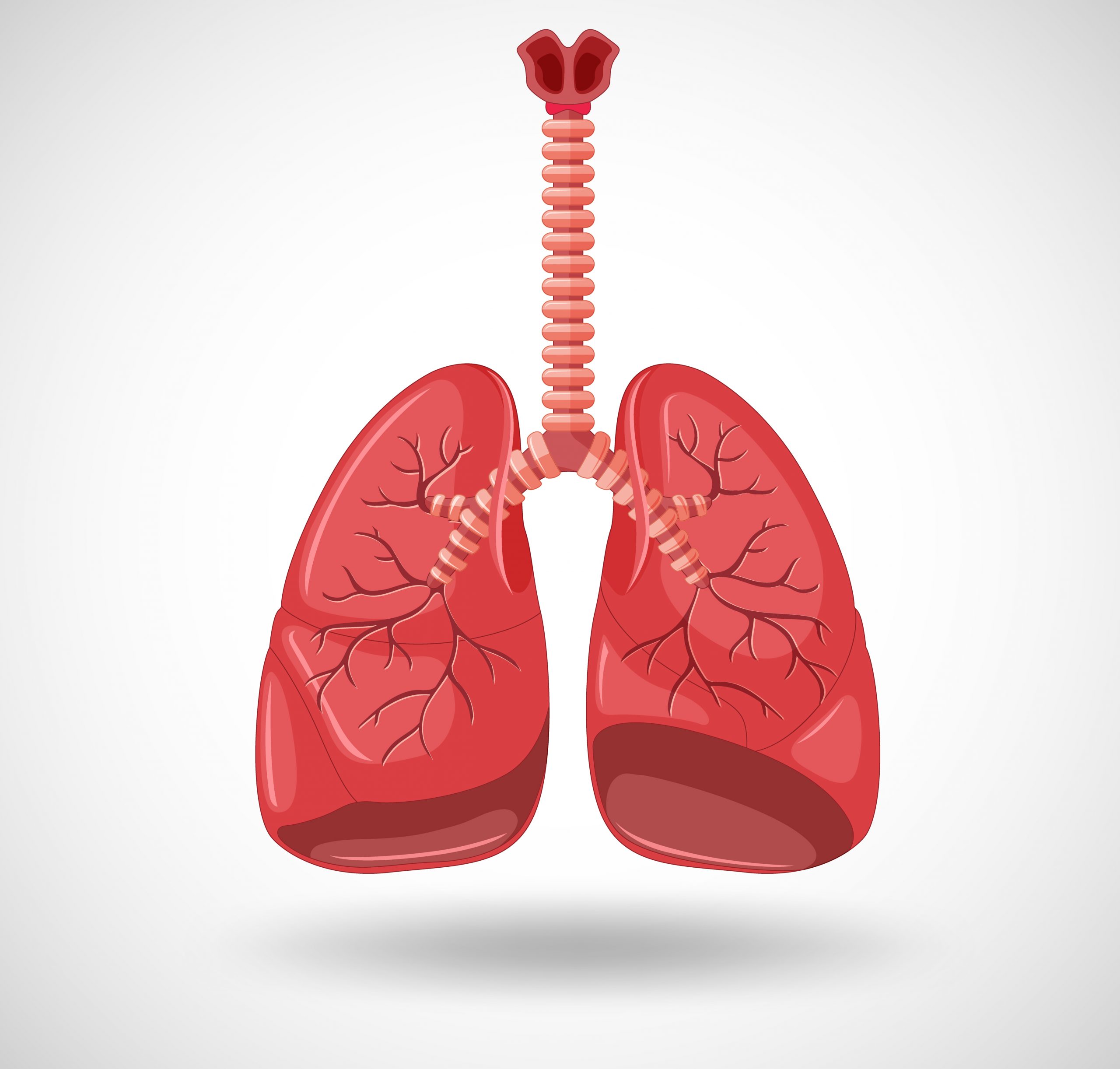

Researchers from the University of Michigan have shown that a form of the rare disease cystinosis is caused by the same cellular mechanism that contributes to a type of cystic fibrosis.
The system eliminates mutant proteins. This enables cystine crystals to accumulate in the cell, a hereditary condition known as cystinosis. As a result, the cell is disrupted, which in turn causes disruption in tissues and ultimately organs—especially the kidneys and the eyes.
When the lysosome, a cell organelle, is unable to carry out its function, a problem arises. The lysosome, often known as the recycling center of the cell, removes waste from the cell and converts it into reusable components that are then transported back into the cell.
However, a cellular mechanism removes the defective protein, causing amino acid, or cystine, to accumulate in the lysosome, when the protein that returns one of the recycled amino acids to the cell mutates and fails.
The Journal of Clinical Investigation has finally published the study’s findings
“If cystinosis not treated at an early age, some of the effects are irreversible and it could include impaired growth, kidney failure and neurological problems,” said Varsha Venkatarangan, graduate student in the U-M Department of Molecular, Cellular and Developmental Biology and lead author of the study.
“Typically, the symptoms of the disease are treated rather than the root problem. So we have been wondering what could be the possible cellular mechanism of this disease.”
Venkatarangan used fibroblasts, which were taken from diseased patients’ skin tissue. She discovered using these cells that the endoplasmic reticulum-associated degradation, or ERAD, disease process destroys a mutant form of the lysosome cystine transporter.
A significant form of cystic fibrosis and other diseases are caused by the same mechanism, ERAD. The discovery of this mechanism also allowed the researchers to demonstrate how previously recognized pharmacological compounds could support the stability of the transporter protein.
“This drug molecule is a small chemical that can somehow assist the protein folding of the mutant and protect it from being degraded,” said MCDB professor Ming Li. “This chemical chaperone helps it achieve the right formation so that it can be functional.”
Li’s laboratory developed an interest in the cystine carrying protein and sought to comprehend how cystinosis was brought on by mutations in the transporter gene. They laboriously gathered roughly 40 cystinosis-causing mutations and investigated the stability of those proteins. One notable mutation was found by the researchers, and compared to the protein’s unaltered version, it had a relatively short half-life.
“We were interested about the fact that this disease mutant was degrading so rapidly, what was the potential mechanism by which it was degrading, and the machinery involved,” Venkatarangan said.
As a result, Venkatarangan conducted a wide range of genetic and chemical studies to inhibit various cell-based protein degradation mechanisms, thereby focusing on the cellular process that was responsible for the condition. ERAD, a biological pathway found in the endoplasmic reticulum of the cell, was identified as the mechanism by the researcher. The endoplasmic reticulum of the cell is a network of minuscule tubes and sheets that aid in transporting proteins throughout the cell. Protein degradation is indicated for misfolded proteins via ERAD.
Drug compounds had already been found to act against the ERAD mechanism and extend the protein’s life because this is a well-known disease mechanism, present in cystic fibrosis. Chemical chaperones are the medicinal molecules that Li referred to as helping the protein form correctly so that it can function.
“This small chemical chaperone can somehow magically—we don’t know the exact mechanism—bind to our mutant and protect the protein from being degraded,” Li said.
After stabilizing the protein and confirming that it was in the proper position to transport amino acids from the lysosome, the researchers had to check to see if the transporter was still active. The researchers determined the amount of cystine in the lysosome by an assay they carried out in association with UCSD. The number of amino acids in the lysosome would be less if the transporter was working properly.
“The reduction was pretty dramatic—nearly a 70% reduction of the accumulation level,” Li said. “That directly brought the system level to almost normal. If we actually used this chemical chaperone to treat patients, it’s possible that we could directly bring down the cystine accumulation in the lysosome.”
People could be perplexed as to why lysosome membrane proteins might be affected by a disease process discovered in the endoplasmic reticulum, Li added. This is so because the endoplasmic reticulum is where lysosome membrane proteins are carried.
There are numerous trafficking proteins required while the lysosome is being constructed. These lysosomal proteins pass via the endoplasmic reticulum before being packaged and sent to the lysosome by the Golgi apparatus. Protein trafficking requires that they fold correctly. Otherwise, protein quality control mechanisms like ERAD will cause them to prematurely breakdown.
The researchers found that these mutant proteins, after becoming trapped and destroyed in the endoplasmic reticulum, vanish early in the trafficking stage.
The researchers also achieved a goal of a National Institutes of Health program that emphasizes finding new uses for already-approved pharmaceuticals as this results in quicker therapies by discovering a new use for an existing drug. The team cautions that the research is preliminary, was only conducted on patient cells grown in culture, and that neither humans nor animals have been tested for or treated with the medicine for cystinosis.
“Again, this is preliminary work that was done in patient cells, so we don’t really know the systemic effect or how well it works in actual patients,” Venkatarangan said. “Our finding also emphasizes the importance of precision medicine, because most of the time, disease mutations are treated similarly under one umbrella of a particular disease.
“But the fact that individual mutations may actually have their own pathogenesis mechanism emphasizes that understanding the individual patient mechanism might help them with a better treatment strategy.”
Weichao Zhang and Xi Yang, both of the U-M MCDB, are co-authors of the research. The study also included contributions from Si Houn Hahn of the Seattle Children’s Hospital at the University of Washington School of Medicine and Jess Thoene of the Department of Pediatrics at the University of Michigan Medical School.
more recommended stories
 AI Predicts Chronic GVHD Risk After Stem Cell Transplant
Key Takeaways A new AI-driven tool,.
 Red Meat Consumption Linked to Higher Diabetes Odds
Key Takeaways Higher intake of total,.
 Pediatric Crohn’s Disease Microbial Signature Identified
Key Points at a Glance NYU.
 Nanovaccine Design Boosts Immune Attack on HPV Tumors
Key Highlights Reconfiguring peptide orientation significantly.
 High-Fat Diets Cause Damage to Metabolic Health
Key Points Takeaways High-fat and ketogenic.
 Acute Ischemic Stroke: New Evidence for Neuroprotection
Key Highlights A Phase III clinical.
 Statins Rarely Cause Side Effects, Large Trials Show
Key Points at a Glance Large.
 Can Too Many Antioxidants Harm Future Offspring?
Key Takeaways High-dose antioxidant supplementation in.
 Anxiety Reduction and Emotional Support on Social Media
Key Summary Anxiety commonly begins in.
 Liquid Biopsy Measures Epigenetic Instability in Cancer
Key Takeaways Johns Hopkins researchers developed.

Leave a Comment