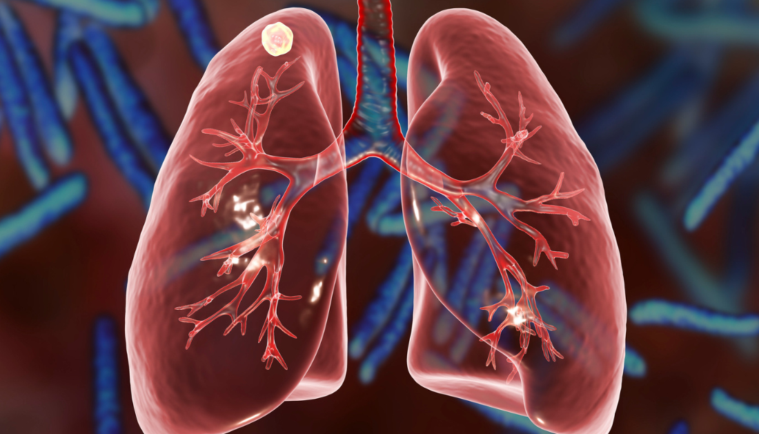

It’s unclear how Mycobacterium TB deflects the immunological response in people, but evidence points to host immunometabolism – the fundamental relationship between immune cell metabolism and immune function. M. tuberculosis is known to impair a metabolic route known as glycolysis in infected myeloid cells, including macrophages, by an unknown method.
A better knowledge of this pathogenic mechanism could lead to a target for the bacterium that will cause 1.6 million deaths in 2021, as well as 10 million new cases of tuberculosis each year.
A new study published in Nature Communications by researchers at the University of Alabama in Birmingham and the Africa Health Research Institute, or AHRI, demonstrates how M. tuberculosis disrupts NADH homeostasis and reprograms glycolysis in myeloid cells. This identifies glycolysis as a potential therapeutic target in the fight against the world’s deadliest infectious illness.
Glycolysis is the process by which glucose is converted into pyruvate while the high-energy molecules ATP and NADH are formed. However, because the system can flow in either direction, researchers were able to use a more specific strategy to limiting glycolytic flux. Previous studies used a sledgehammer approach, such as applying an inhibitor that limits glucose uptake into myeloid cells.
The enzyme lactate dehydrogenase, abbreviated LDH, catalyzes the reversible process of lactate fermentation. LDH is made up of four subunits, which are a mix of LDHA and LDHB subunits. When LDH is composed primarily of LDHA subunits, which are abundant in myeloid cells, it preferentially converts pyruvate to lactate and NADH to NAD+. An LDH composed of LDHB subunits, on the other hand, favors the opposite reaction.
LHDA’s significance in TB pathogenesis is unknown. The UAB and AHRI researchers examined resected lung tissue from tuberculosis patients and discovered that myeloid, bronchial epithelial cells, and lymphocytes stained positive for LDHA while engaging in unique immunological processes such as granuloma formation and alveolitis. “These findings point to LDHA as an important metabolic protein in the immune response in human tuberculosis lesions,” stated senior author Adrie Steyn, Ph.D.
Steyn and colleagues proposed that NAD(H)-mediated glycolytic flux in myeloid cells protects the host from M. tuberculosis infection because NADH/NAD+ regulates glycolysis at defined phases. They generated mice lacking the LDHA component in myeloid cells to test this. These cells have a lower glycolytic capacity because the altered LDH activity, which is made up entirely of LDHB subunits, reduces their ability to produce NAD+ from NADH in the presence of pyruvate.
When infected with a low dosage of M. tuberculosis, the LDHA-deficient mice were more vulnerable to infection and had a considerably shorter survival period. In addition, the LDHA-deficient animals had poorer gross pathology and histology of the lungs. Furthermore, wild-type mice produce a robust inflammatory response as a protective immunological response to M. tuberculosis infection, but LDHA-deficient mice show little early inflammation.
“This suggests that LDHA is necessary for protection against tuberculosis and that glycolytic flux in myeloid cells is essential for the control of M. tuberculosis infection and disease,” he said.
Despite signs of a diminished immune response, researchers discovered that when they assessed gene expression in the lungs of LDHA-deficient mice, mRNAs linked with inflammatory processes were among the most enriched, particularly a robust interferon-gamma gene set. “The robust interferon-gamma gene expression signature in more susceptible mice with a blunted immune response was particularly intriguing since interferon-gamma is an indispensable antimycobacterial cytokine widely considered to be protective in tuberculosis,” Steyn said in a statement.
Bioenergetics tests revealed that mouse macrophages require LDHA and its LDH-mediated NAD+ regeneration for their metabolic response to interferon-gamma.
Because NAD+ depletion appeared to be important to M. tuberculosis’ glycolytic suppression, the researchers wondered if adding a NAD+ precursor, nicotinamide, might modify macrophages’ ability to generate an immunological response.
Nicotinamide was discovered to boost the glycolytic capacity of M. tuberculosis-infected bone marrow macrophages. The researchers postulated that nicotinamide works as a host-directed treatment by increasing glycolysis in M. tuberculosis-infected macrophages via the NAD+ salvage pathway.
In vitro trials in which they infected macrophages with luciferase-expressing M. tuberculosis revealed that nicotinamide was an effective tuberculosis treatment. Nicotinamide reduced luminescence in infected macrophages 48 hours after infection in a dose-dependent manner, and this reduction of pathogenic bacteria was dependent on glycolysis. In a mouse model, feeding the mice nicotinamide for four weeks, beginning three days or 28 days after infection, resulted in a tenfold reduction of M. tuberculosis load in the lungs, as well as a reduction in inflammation.
Nicotinamide was first described as a tuberculosis treatment in the 1940s, via a different mechanism; nevertheless, it was mostly abandoned when much more efficient medications were discovered during the antibiotic golden age.
However, the tuberculosis environment has altered considerably in the previous 60 years. According to Steyn, the incidence of tuberculosis has risen to more than 10 million new cases per year, and the disease has gained resistance to the frontline medications that have replaced nicotinamide.
“We have provided further evidence of a host-dependent effect of nicotinamide, the metabolic requirements for its activity and a modern-day demonstration of its efficacy as a treatment for tuberculosis, using two treatment regimens in vivo,” Steyn remarked in regards to the current research. “Logistically, nicotinamide meets many of the World Health Organization’s criteria for an optimal novel tuberculosis treatment regimen.” It is affordable, orally accessible, shelf-stable, incredibly safe and acceptable, and widely used in humans for a variety of purposes. Finally, these properties make nicotinamide intriguing as an old tool in a modern context.”
Steyn claims that there is still an unsolved question. M. tuberculosis depletes NAD(H) levels in what way? According to the researchers, one possible explanation is that M. tuberculosis secretes tuberculosis necrotizing toxin, or TNT, a NAD+ glycohydrolase. This toxin, discovered in 2015 by UAB’s Michael Niederweis, Ph.D., is the first toxin discovered in M. tuberculosis in 132 years of research. TNT dramatically lowers NAD+ abundance in infected macrophages in wildtype M. tuberculosis.
Steyn is a UAB professor of microbiology who directs labs at UAB and the AHRI in Durban, KwaZulu Natal, South Africa, the world’s epicenter for tuberculosis infections. Niederweis is a microbiology professor at the University of Alabama at Birmingham.
Hayden T. Pacl, M.D., Ph.D., of the UAB Department of Microbiology, is the first author of the article, “NAD(H) homeostasis underpins host protection mediated by glycolytic myeloid cells in tuberculosis.”
For more information : NAD(H) homeostasis underlies host protection mediated by glycolytic myeloid cells in tuberculosis, Nature Journal
more recommended stories
 Acute Ischemic Stroke: New Evidence for Neuroprotection
Key Highlights A Phase III clinical.
 Statins Rarely Cause Side Effects, Large Trials Show
Key Points at a Glance Large.
 Anxiety Reduction and Emotional Support on Social Media
Key Summary Anxiety commonly begins in.
 Liquid Biopsy Measures Epigenetic Instability in Cancer
Key Takeaways Johns Hopkins researchers developed.
 Human Antibody Drug Response Prediction Gets an Upgrade
Key Takeaways A new humanized antibody.
 Pancreatic Cancer Research: Triple-Drug Therapy Success
Key Summary Spanish researchers report complete.
 Immune Cell Epigenome Links Genetics and Life Experience
Key Takeaway Summary Immune cell responses.
 Dietary Melatonin Linked to Depression Risk: New Study
Key Summary Cross-sectional analysis of 8,320.
 Chronic Pain Linked to CGIC Brain Circuit, Study Finds
Key Takeaways University of Colorado Boulder.
 New Insights Into Immune-Driven Heart Failure Progression
Key Highlights (Quick Summary) Progressive Heart.

Leave a Comment