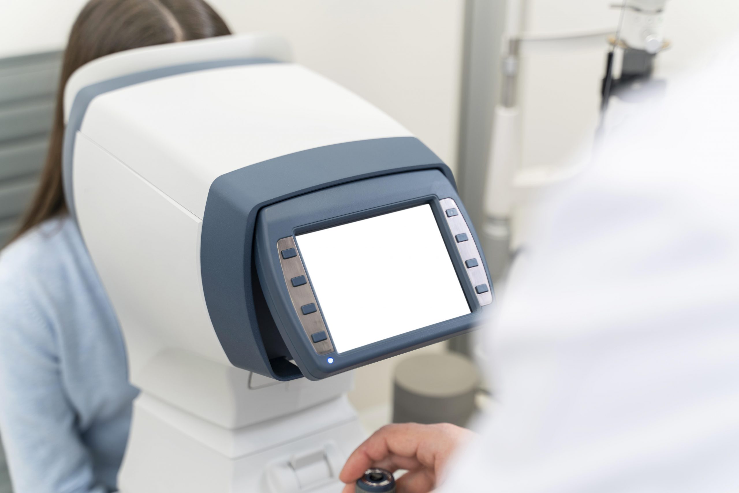

Using retinal pictures from the eye, Duke Health researchers developed a machine learning model that can distinguish between normal cognition and mild cognitive impairment.
To identify people with mild cognitive impairment, the model analyzes retinal pictures and accompanying data and recognizes particular traits. The model, which was published in the journal Ophthalmology Science, shows the possibility for a non-invasive and low-cost technique of detecting early indicators of cognitive impairment that could lead to Alzheimer’s disease.
“This is particularly exciting work because we have previously been unable to differentiate mild cognitive impairment from normal cognition in previous models,” said senior author Sharon Fekrat, M.D., professor in Duke’s departments of Ophthalmology and Neurology, and associate professor in the Department of Surgery. “This work brings us one step closer to detecting cognitive impairment earlier before it progresses to Alzheimer’s dementia.”
Fekrat and colleagues previously built a model that successfully identified patients with a known Alzheimer’s diagnosis using retinal scans and other data. The scans, which used optical coherence tomography (OCT) and optical coherence tomography angiography (OCTA), identified structural alterations in the neurosensory retina and its microvasculature in Alzheimer’s patients.
The present study builds on prior research by employing machine learning techniques to detect moderate cognitive impairment, which is frequently a precursor to Alzheimer’s disease. The new model recognizes certain elements in OCT and OCTA images that indicate the existence of cognitive impairment, as well as patient variables such as age, gender, visual acuity, and years of schooling, as well as quantitative data from the images themselves.
The algorithm evaluated retinal pictures and images, as well as quantitative data, to distinguish those with normal cognition from those with a diagnosis of mild cognitive impairment, with a sensitivity of 79% and specificity of 83%, according to the researchers.
“This is the first study to use retinal OCT and OCTA images to distinguish people with mild cognitive impairment from individuals with normal cognition,” said co-first author C. Ellis Wisely, M.D., assistant professor in the Department of Ophthalmology.
“Having a non-invasive and less expensive means to reliably identify these patients is increasingly important, particularly as new therapies for Alzheimer’s disease may become available,” Wisely said.
“The retina is a window to the brain, and machine learning algorithms that leverage non-invasive and cost-effective retinal imaging to assess neurological health can be a potent tool to screen patients at scale,” said co-lead author Alexander Richardson, a student in the Eye Multimodal Imaging in Neurodegenerative Disease lab at Duke.
more recommended stories
 Nanoplastics in Brain Tissue and Neurological Risk
Key Takeaways for HCPs Nanoplastics are.
 AI Predicts Chronic GVHD Risk After Stem Cell Transplant
Key Takeaways A new AI-driven tool,.
 Red Meat Consumption Linked to Higher Diabetes Odds
Key Takeaways Higher intake of total,.
 Pediatric Crohn’s Disease Microbial Signature Identified
Key Points at a Glance NYU.
 Nanovaccine Design Boosts Immune Attack on HPV Tumors
Key Highlights Reconfiguring peptide orientation significantly.
 High-Fat Diets Cause Damage to Metabolic Health
Key Points Takeaways High-fat and ketogenic.
 Acute Ischemic Stroke: New Evidence for Neuroprotection
Key Highlights A Phase III clinical.
 Statins Rarely Cause Side Effects, Large Trials Show
Key Points at a Glance Large.
 Can Too Many Antioxidants Harm Future Offspring?
Key Takeaways High-dose antioxidant supplementation in.
 Anxiety Reduction and Emotional Support on Social Media
Key Summary Anxiety commonly begins in.

Leave a Comment