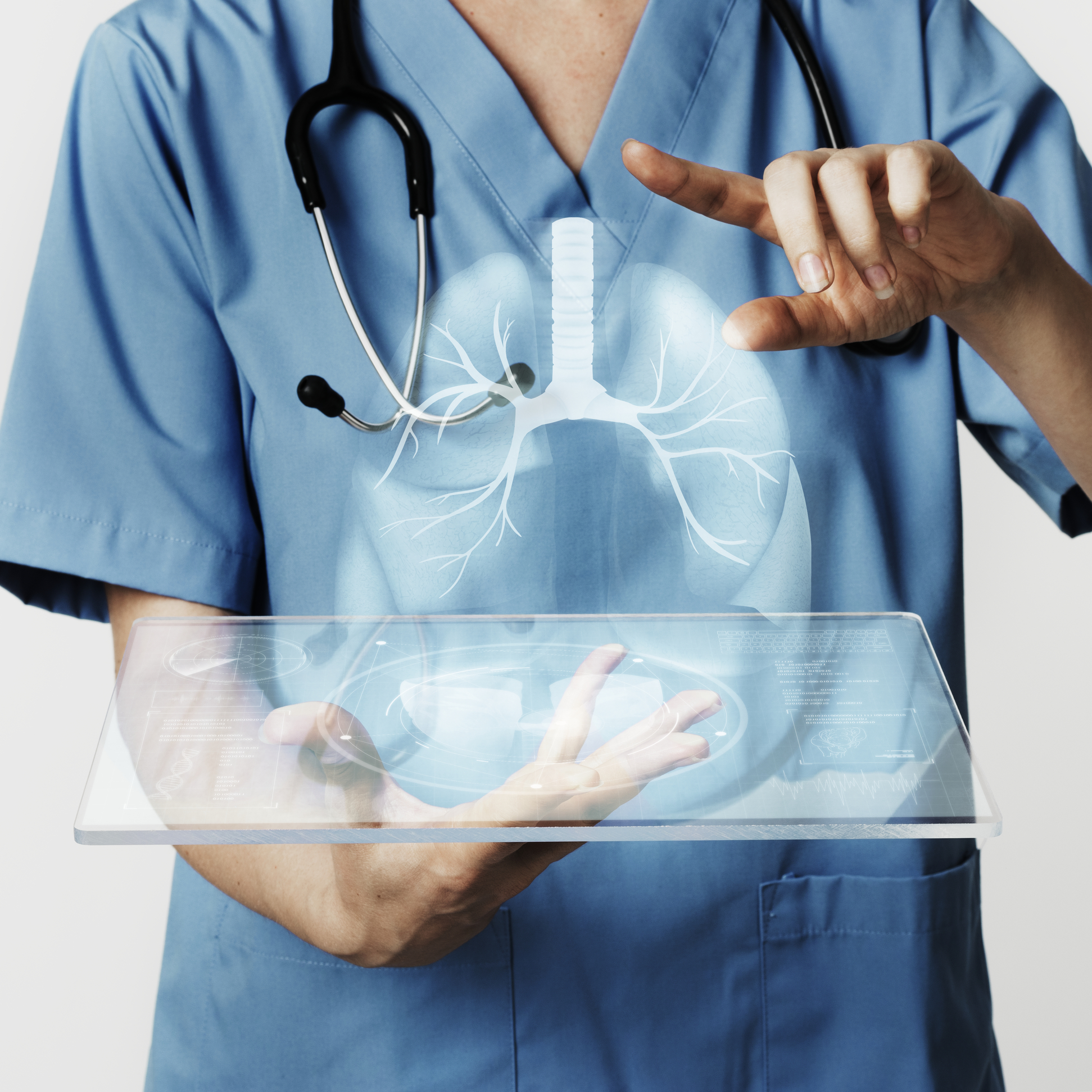
Image by rawpixel.com on Freepik
Scientists developed a blueprint for mini-lungs that will accelerate the discovery and development of new drugs and reduce the reliance on animal testing. When we’re going to a new location, we frequently turn down the audio as we follow the directions. What had been a melody now sounds like noise, interfering with our concentration.
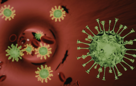
Noise has similarly complicated our understanding of how viral diseases such as COVID affect human lungs. The data from patient lung tissues varies widely from person to person, confounding the core mechanics of how SARS-CoV-2 first infects lung cells. It’s also an after-the-fact study, as if we’re trying to trace the virus’s path three states back.
Turning down the noise of diversity by analyzing genetically similar tissues from the moment of infection could shed light on the pathogen’s journey. When and which cells become infected? What is the infection level, and how does it differ based on cell type? How does it alter under different conditions?
What if it was able to track thousands of these illnesses at once? It has the potential to change our understanding of both infections and the drugs used to treat them.
That is the goal for more advanced technology capable of producing small organs on microchips. Ali Brivanlou and Charles M. Rice of Rockefeller University cooperated to create a cell culture technology platform that generates genetically identical lung buds—the embryonic structures that give rise to our breathing organs—from human embryonic stem cells (hESCs). The findings were recently reported in Stem Cell Reports.
When placed on a microchip array and carefully dosed with a particular cocktail of signaling molecules, the hESCs swiftly organize themselves into “mini-lungs” with complete tissue complexity. These buds can be cultivated in the hundreds, enabling for unparalleled high-throughput investigation of lung tissue infection without all the noise variables.
As a result, there is unrestricted, rapid, and scalable access to lung tissue that exhibits the main characteristics of human lung development and may be utilized to follow lung infections and identify candidate therapies.
“These lungs are basically clones,” says Ali Brivanlou. “They have the exact same DNA signature. That way we don’t have to worry about one patient responding differently from another. Quantification allows us to keep the genetic information constant and measure the key variable—the virus.”
Creating a More Effective Mini-Lungs
Embryonic stem cells can divide indefinitely to produce additional stem cells or to develop into any tissue. Brivanlou’s Laboratory of Synthetic Biology has long investigated their possibilities.
During the COVID pandemic, Brivanlou collaborated with Rockefeller colleague Charles M. Rice: his lab had the microchip technology to produce lung buds, and Rice’s lab had the requisite biosafety clearance to infect them with SARS-CoV-2 and evaluate the outcome.
Edwin Rosado-Olivieri, a stem cell biologist in Brivanlou’s lab, and Brandon Razooky, then a postdoc in Rice’s Laboratory of Virology and Infectious Disease, started coaxing the cells to organize into more specialized forms in 2021. Stem cells do not spontaneously assemble. They require a limited location, such as a microchip well, as well as stimuli to cause change. The stimuli originate from four major signaling pathways, which cause stem cells to develop into certain cell types.
After around two weeks, the lung cells in the group generated similar buds with molecular profiles that nearly mirrored those seen in the first stages of fetal lung development, including the production of airways and alveoli, features known to be disrupted in many persons with severe COVID.
Identifying the Main Culprit
Since then, the platform has been utilized by researchers to better understand how SARS-CoV-2 infects distinct lung cells.
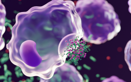
Alveoli are small sacs at the ends of lung branches that control the gas exchange that occurs with each breath: oxygen in, carbon dioxide out. The researchers determined that alveoli were more susceptible to SARS-CoV-2 infection than airway cells, which are the organ’s guardians and the first line of defense against all inhaled threats by analyzing cloned alveolar cells in large numbers. If the virus got past them, the alveoli were dead meat.
They also discovered a winning signaling protein combination for producing the most robust batches of lung buds: a combination of keratinocyte growth factor (KGF) and bone morphogenetic protein 4 (BMP4). Both aid in cell differentiation and expansion.
Surprisingly, the BMP route has a disadvantage. When the researchers compared infected lung buds to COVID patients’ post-mortem tissue, they discovered that the BMP signaling pathway was activated in both, making the tissues more susceptible to infection. By blocking the BMP pathway, the cells became less susceptible.
Apart from COVID
The platform can also be used to explore the mechanisms of influenza, RSV, respiratory disorders, and lung cancer, among other ailments, according to the researchers. It can also be used to screen for new medications to treat them.
 Transcatheter Valve-in-Valve Improves Mitral Outcomes
Key Highlights Transcatheter mitral valve-in-valve (mVIV).
 Stroke Rehabilitation: Early High-Intensity Therapy Findings
Key Highlights High-intensity therapy within 2.
 TRPM8 Cold Sensation Mechanism Explained for Pain Care
Quick Summary TRPM8 ion channel converts.
 Gum Recession from Snus Confirmed, Caries Risk Debated
Key Highlights Snus use is strongly.
 Hypertensive Disorders of Pregnancy: Role of Daily Activity
Key Points Summary Limiting sedentary time.
 Climate Change Drives Dengue Outbreaks Globally
Key Takeaways Extreme weather significantly increases.
 Teen Driving Risks: Parents Underestimate Safety Threats
Key Takeaways Teen driving risks remain.
 PFK Enzyme Dual Role in Metabolism and Cell Cycle
Key Highlights Phosphofructokinase (PFK Enzyme) shows.
 Exercise During Chemotherapy Supports Brain Health
Key Points Summary A nationwide clinical.
 Invasive Cosmetic Procedures Raise Patient Safety Concerns
Key Summary Experts publishing in The.
Leave a Comment