Image by brgfx on Freepik
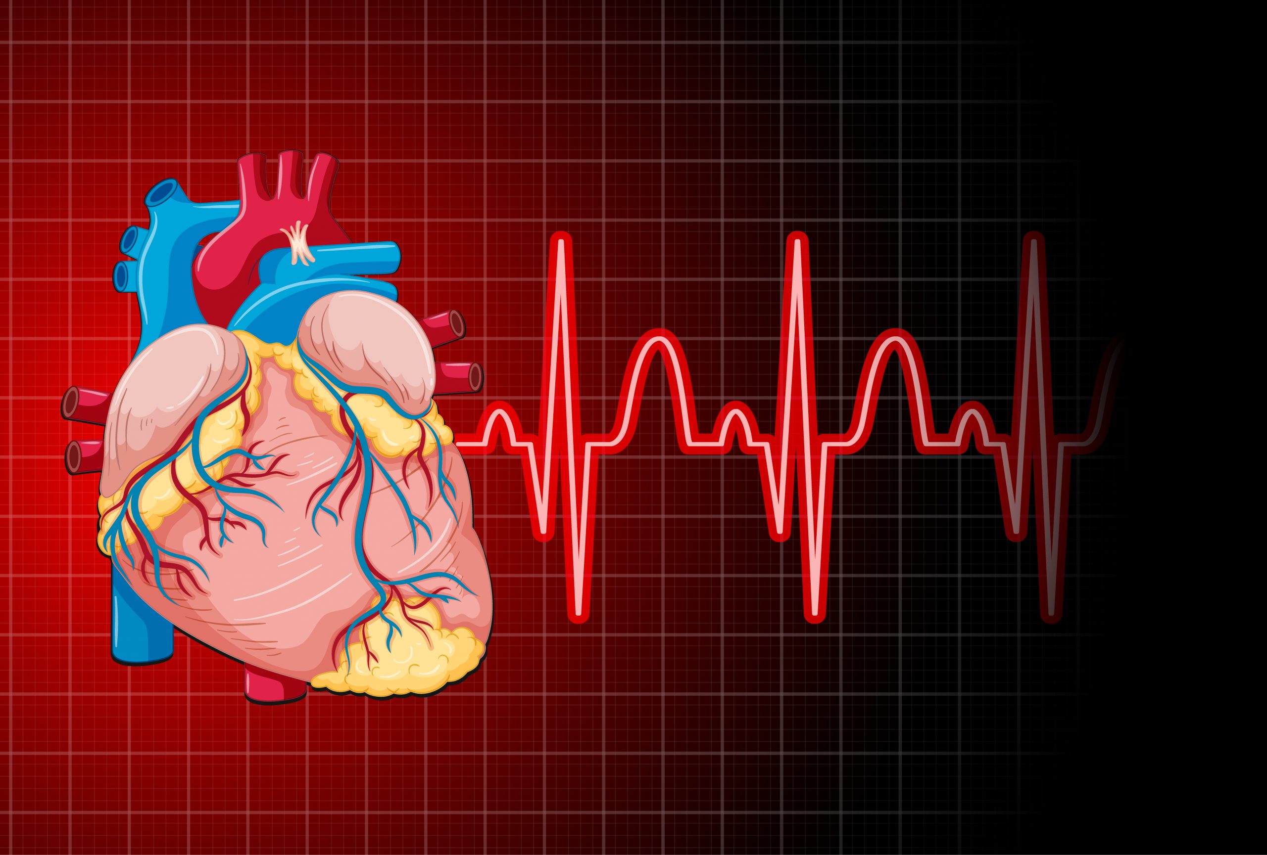
Researchers from King’s College London devised and validated the first end-to-end pipeline for fully automated analysis of huge, unstructured clinical and research database of cardiac magnetic resonance- CMR scan.
Many hospitals have huge CMR databases that, when linked with electronic health records, can provide important insights into treatment efficacy and shape future healthcare research and guidelines.
However, databases are frequently organized differently by different organizations and may contain missing or duplicated files, posing a considerable obstacle for data processing.
Previously, human curation and analysis by healthcare specialists would have needed a major time investment, whereas an AI tool can be taught to efficiently ‘data wrangle’ at scale, quickly assess quality, and translate data into standard forms and structures.
Researchers from the School of Biomedical Engineering & Imaging Sciences trained a generalizable AI algorithm on data from over 7000 CMR scans, with preliminary results demonstrating human-level accuracy for left ventricle and right ventricle segmentations across all major CMR scanner technologies, as well as for a wide range of cardiac diseases.
“The proposed framework is a fundamental step for the clinical translation of AI algorithms. In addition, it allows for retrospective analysis of large clinical (research) datasets and compared to other works it allows analysis of a much larger set of biomarkers for regional and global systolic and diastolic biventricular function from CMR scans.” – Dr Esther Puyol, Visiting Lecturer, Department of Biomedical Engineering
“This work addresses the under-exploitation of the large clinical CMR imaging databases that many hospitals worldwide maintain. These are incredibly rich resources, containing historical and longitudinal data from many thousands of patients. When combined with data from electronic health records they could provide valuable insight into the effectiveness of treatments to inform future guidelines.” – Dr Andrew King, Reader in Medical Image Analysis, Department of Biomedical Engineering
Future research will aim to extend this paradigm in order to identify biomarkers associated with systolic and diastolic function, as well as their potential links to patient therapies and healthcare outcomes.
 Breast Cancer Risk Rises with Aging Tissue Changes
Key Highlights A 3-million-cell atlas reveals.
 Type 2 Diabetes Risk Rising in Genetically Susceptible
Quick Summary Rising type 2 diabetes.
 Female Microbiome Shaped by Diet, Stress, Unhealthy Lifestyle
Quick Summary Lifestyle factors significantly influence.
 Transcatheter Valve-in-Valve Improves Mitral Outcomes
Key Highlights Transcatheter mitral valve-in-valve (mVIV).
 Stroke Rehabilitation: Early High-Intensity Therapy Findings
Key Highlights High-intensity therapy within 2.
 TRPM8 Cold Sensation Mechanism Explained for Pain Care
Quick Summary TRPM8 ion channel converts.
 Gum Recession from Snus Confirmed, Caries Risk Debated
Key Highlights Snus use is strongly.
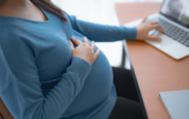 Hypertensive Disorders of Pregnancy: Role of Daily Activity
Key Points Summary Limiting sedentary time.
 Climate Change Drives Dengue Outbreaks Globally
Key Takeaways Extreme weather significantly increases.
 Teen Driving Risks: Parents Underestimate Safety Threats
Key Takeaways Teen driving risks remain.
Leave a Comment