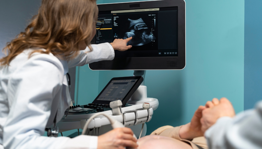

Approximately 10% of pregnant newborns are classified as undersized for their gestational age. If these babies are still healthy, no intervention is required during pregnancy. However, in the event of small babies with a dysfunctional placenta, intervention is required, and the baby’s birth may need to be induced.
“This means it is incredibly important to track down which babies are smaller due to the placenta,” says Wessel Ganzevoort, associate professor of obstetrics at Amsterdam UMC and study leader.
Doppler measurements
For nearly 50 years, growth ultrasounds have been used to detect small newborns and determine whether they follow their own growth pattern or begin to grow more slowly. In this study, little newborns received a Doppler ultrasonography in addition to the conventional growth measurement. This ultrasound analyzes the resistance of the blood arteries in the umbilical cord, revealing information regarding blood flow to the placenta.
The ultrasound can also detect blood flow to the child’s brain. If the supply is higher than usual, it could indicate that the placenta isn’t functioning properly. The baby has then “opened” a blood artery in the brain to protect it from the shortfall created by a failing placenta. With a poor placenta, the kid is more likely to experience health problems (such as a lack of oxygen) and, eventually, death at birth.
The study also looked into whether the child’s results improved if the delivery was induced before 37 weeks of gestation. This did not result in better outcomes. The recommendation is to wait until at least 37 weeks of pregnancy to induce labor. This is because it is preferable for the kid to remain in the womb for as long as possible, provided there are no additional concerns of health issues.
Added Value
“What was achievable with a Doppler ultrasonography was long recognized, but it is still not standard practice at all hospitals. This study now demonstrates that this measurement has extra value for diagnosing pregnancies in newborns that are too small and have a faulty placenta,” explains Mauritia Marijnen, a Ph.D. candidate at Amsterdam UMC and study’s first author.
“By incorporating this Doppler ultrasound into the care plan for these small newborns, the increased risk of complications during labor can be effectively identified and managed. Small newborns with appropriate measurements can be watched less closely. As a result, Ganzevoort finds that the delivery is more likely to occur naturally and without intervention.
For more information: Doppler ultrasound of umbilical and middle cerebral artery in third trimester small for gestational age fetuses to decide on timing of delivery for suspected fetal growth restriction: a cohort with nested RCT (DRIGITAT), British Journal of Obstetrics and Gynaecology (2024), https://doi.org/10.1111/1471-0528.17770
more recommended stories
 Phage Therapy Study Reveals RNA-Based Infection Control
Key Takeaways (Quick Summary) Researchers uncovered.
 Safer Allogeneic Stem Cell Transplants with Treg Therapy
A new preclinical study from the.
 AI in Emergency Medicine and Clinician Decision Accuracy
Emergency teams rely on rapid, accurate.
 Innovative AI Boosts Epilepsy Seizure Prediction by 44%
Transforming Seizure Prediction in Epilepsy Seizure.
 Hypnosis Boosts NIV Tolerance in Respiratory Failure
A New Approach: Hypnosis Improves NIV.
 Bee-Sting Microneedle Patch for Painless Drug Delivery
Microneedle Patch: A Pain-Free Alternative for.
 AI Reshapes Anticoagulation in Atrial Fibrillation Care
Understanding the Challenge of Atrial Fibrillation.
 Hemoglobin as Brain Antioxidant in Neurodegenerative Disease
Uncovering the Brain’s Own Defense Against.
 Global Data Resource for Progressive MS Research (Multiple Sclerosis)
The International Progressive MS Alliance has.
 AI Diabetes Risk Detection: Early T2D Prediction
A new frontier in early diabetes.

Leave a Comment