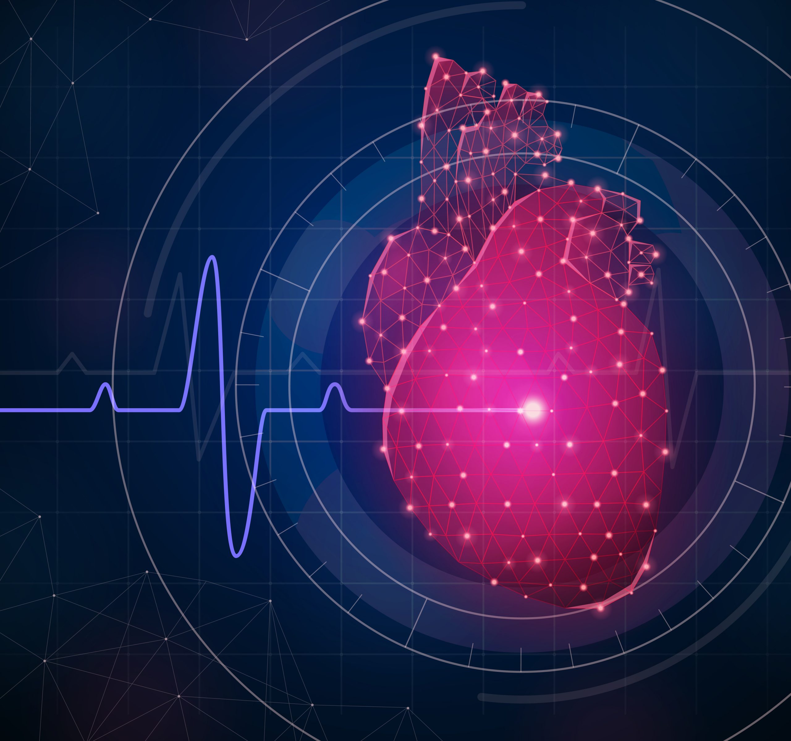

In a groundbreaking achievement, scientists from the Cardiovascular Data Science (CarDS) Lab have introduced an innovative method capable of identifying a prevalent valvular heart condition, recognized as severe aortic stenosis, through echocardiograms. This significant research, unveiled on August 23 in the European Heart Journal, carries the potential to revolutionize standard clinical practices.
Severe aortic stenosis (AS), a prominent health concern among older adults due to a narrowed aortic valve, prompts the need for timely diagnosis. This aids in curbing symptoms and minimizing the risk of untimely death and hospitalization.
Doppler echocardiography, a specialized ultrasound heart imaging, stands as the primary diagnostic tool for AS detection. The team has ingeniously devised a deep learning model capable of utilizing simplified heart ultrasound scans for automated identification of severe AS.
Leading this technological advancement is Rohan Khera, MD, MS, an adept figure in cardiovascular medicine and health informatics. As an assistant professor and director of the CarDS Lab, Khera collaborated with colleagues from the Chandra Family Department of Electrical and Computer Engineering at UT Austin. The study encompassed an impressive 5,257 studies, including 17,570 videos spanning 2016 to 2020 at Yale New Haven Hospital. This model’s efficacy was subsequently confirmed through a comprehensive external validation from 2,040 consecutive studies from distinct cohorts in New England and California.
“Our challenge is that precise evaluation of AS is crucial for patient management and risk reduction. While specialized testing remains the gold standard, reliance on those who make it to our echocardiographic laboratories likely misses people early in their disease state,” said Khera.
“Our goal was to develop a machine learning approach that would be suitable for point-of-care ultrasound screening,” said the study’s co-first author Evangelos Oikonomou, MD, DPhil, a cardiology fellow and a current postdoctoral researcher in the CarDS Lab.
Through their efforts, the possibility for identifying aortic stenosis at an early stage emerges, paving the way for prompt patient intervention. “Our work can allow broader community screening for AS as handheld ultrasounds can increasingly be used without the need for more specialized equipment. They are already being used frequently in emergency departments, and many other care settings,” added Khera.
This achievement emerges as a product of harmonious cooperation between clinician-researchers and computer experts. Notably, the instrumental role played by Greg Holste, a doctoral candidate at UT Austin, under the joint guidance of Dr. Khera, stands out. Holste took charge of devising a pioneering approach that facilitated the technology, simultaneously earning the status of a co-first author alongside Dr. Khera in the study’s development.
more recommended stories
 Liquid Biopsy Measures Epigenetic Instability in Cancer
Key Takeaways Johns Hopkins researchers developed.
 Human Antibody Drug Response Prediction Gets an Upgrade
Key Takeaways A new humanized antibody.
 Pancreatic Cancer Research: Triple-Drug Therapy Success
Key Summary Spanish researchers report complete.
 Immune Cell Epigenome Links Genetics and Life Experience
Key Takeaway Summary Immune cell responses.
 Dietary Melatonin Linked to Depression Risk: New Study
Key Summary Cross-sectional analysis of 8,320.
 Chronic Pain Linked to CGIC Brain Circuit, Study Finds
Key Takeaways University of Colorado Boulder.
 New Insights Into Immune-Driven Heart Failure Progression
Key Highlights (Quick Summary) Progressive Heart.
 Microplastic Exposure and Parkinson’s Disease Risk
Key Takeaways Microplastics and nanoplastics (MPs/NPs).
 Sickle Cell Gene Therapy Access Expands Globally
Key Summary Caring Cross and Boston.
 Reducing Alcohol Consumption Could Lower Cancer Deaths
Key Takeaways (At a Glance) Long-term.

Leave a Comment