Image by macrovector on Freepik
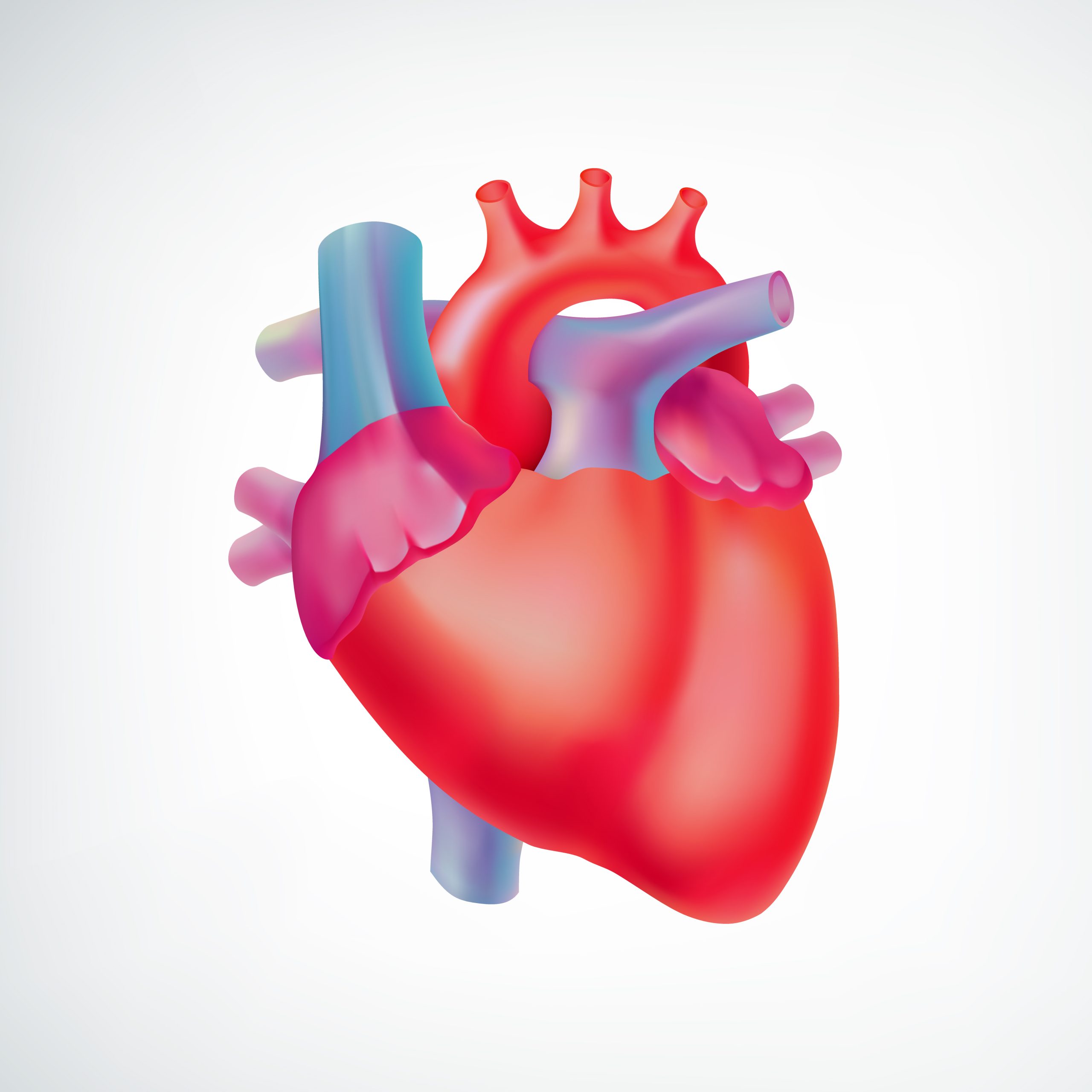
If you’ve ever attempted to photograph a puppy, the result was probably just a blur of fur. Consider the difficulty researchers who are trying to understand the electrical conductivity of heart muscle must overcome when trying to read a stock ticker on a puppy’s fur.
Every second, the beating heart tissue contracts and relaxes as it pumps, but for scientists researching electrical conduction, this vital function is a confusing one they refer to as a “motion artifact.” Researchers must employ medications to stop the heart cells from beating while they collect their measurements in order to interpret the stock ticker, or the moving fluorescent signals that show how electrically the heart cells are operating. This may have an impact on cardiac electrophysiology and make it more difficult to understand how electrical conduction is related to cellular structure and mechanical contraction.
An innovative computational technique has been created by Washington University in St. Louis researchers to eliminate motion in pictures of expanding and contracting cardiac cells and tissues. The algorithm imitates a drug’s function in stopping the heart by computationally eliminating movement, while maintaining cellular structure and tissue contractility.
The findings of the study, which was directed by Guy Genin, the Harold and Kathleen Faught Professor of Mechanical Engineering, and Nathaniel Huebsch, an assistant professor of biomedical engineering at the McKelvey School of Engineering, were published in the Proceedings of the National Academy of Sciences.
“When the heart beats, the electrical signal goes through the tissue, and that triggers mechanical squeezing,” said Huebsch, who studies how mechanical cues affect heart development and disease.
“In many cases, deadly arrhythmias happen because the electric pulse becomes decoupled from the mechanical squeezing. Our group leverages biomaterials and genetics to study how inherited cardiomyopathies develop, and we are very excited to have a tool that allows us, for the first time, to directly and non-invasively study electro-mechanical coupling in cardiomyocytes,” Huebsch continued.
The team’s algorithm imitates the effects of the drug blebbistatin, which slows down heartbeats while simultaneously having an adverse effect on the cells’ basic structure. Eliot Elson, Emeritus Professor of Biochemistry and Molecular Biophysics at the School of Medicine, collaborated with Genin and Huebsch on the technique.
Louis Woodhams, senior lecturer in mechanical engineering & materials science and a McKelvey Engineering alumnus who earned a master’s and a doctorate in 2017 and 2022, and Jingxuan Guo, who obtained a doctorate in mechanical engineering & materials science in 2022, collaborated to develop the technique and refine it for use in heart tissues created from human induced pluripotent stem cells (iPSCs).
In order to transfer developing signals back to a stabilized image of the tissue, two algorithms were used to construct virtual blebbistatin: one that has been successful in calculating displacements for cardiac mapping, and the other that leverages the first. Huebsch claimed that this strategy allowed the researchers to directly monitor the coupling of calcium waves, membrane voltage, and mechanical contraction beat by beat in his lab’s iPSC heart tissue models, giving them insight into heart diseases. Virtual blebbistatin does not impair tissue contractility, he added.
Hypertrophic cardiomyopathy, a hereditary illness in which the heart muscle thickens and stiffens, making it harder for the heart to beat, is one of those diseases. The most frequent natural cause of mortality in children and young adults, the condition is frequently quiet and is discovered only when a young athlete passes away unexpectedly during competition. It affects roughly one in 500 persons.
“Not everybody who has the gene mutation will develop the disease, which makes it really hard to manage the treatment of the patients,” Huebsch said. “Normally, if someone is at high risk to develop deadly arrhythmias, you’d want to implant a pacemaker in them. But since not everyone with these mutations will develop the disease, that’s not a good option. On top of this challenge, studies on new drugs to treat the disease require treatment to begin very early on in the course of disease in controlled animal models.
The group used synthetic heart muscle as a model and tested virtual blebbistatin on cardiomyocytes made from human iPSCs. In Elson’s laboratory, the first synthetic cardiac tissues were created in the 1990s. The tissue models developed by Huebsch’s team expand on this by using patient-specific cardiac cells that can be produced from a patient’s fat cells in surroundings that are dynamic and elastic to mimic disease and development. is starting to identify which people are at risk of having severe cardiac disease utilizing the most up-to-date genomic technologies to comprehend how genes interact with epigenetic variables.
In the study, scientists compared genetically modified heart cells for hypertrophic cardiomyopathy to heart cells with designed heart muscle, and they were able to assess mechanical-electrical coupling in the two by removing around 95% of motion from them.
“The electrical-mechanical coupling of heart tissue is a key factor in the mechanobiology of heart function,” Genin said. This combination of the Huebsch lab’s advances in laboratory disease modeling with the latest solid mechanics tools provides much potential for understanding how epigenetic factors affect the mechanobiology of disease progression.
 Calcific Aortic Valve Stenosis Linked to Gum Disease Bacteria
Key Points Researchers identified a potential.
 Drug Delivery Nanoparticles Studied with New AF4-SANS Tool
Key Points Researchers developed the first.
 Gut-Friendly Diet Linked to Lower CHD Mortality Risk
Key Summary A higher Dietary Index.
 Parkinson’s Disease Risk Linked to Short Sleep, Study Finds
Key Points A large Chinese cohort.
 Cancer Risk And Allergic Diseases Linked in New Study
Key Points A large meta-analysis of.
 Lifestyle Behaviors and Mood Linked in Daily Life Study
Key Points A 70-day diary study.
 Cat Fleas Linked to Murine Typhus Risk in Texas Study
Key Points Researchers detected Rickettsia typhi.
 Pediatric Surgical Safety Reduces Serious Surgical Events
Key Points Pediatric operating room safety.
 England Obesity Prevalence Climbs to 30%, Lancet Study
Key Points A nationwide study of.
 Rheumatoid Arthritis Linked to SPP1hi Macrophages Growth
Key Summary Researchers at Hospital for.
Leave a Comment