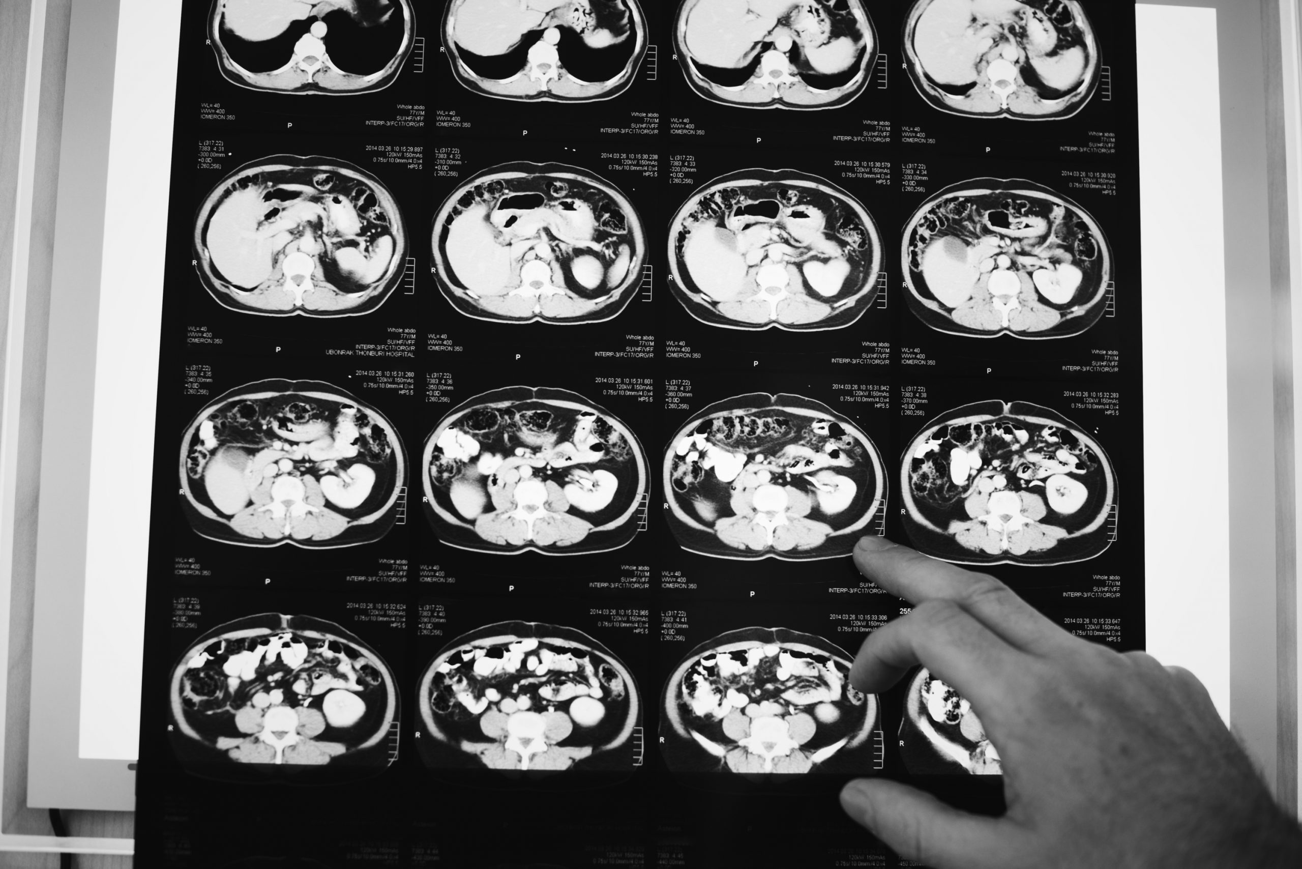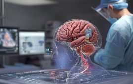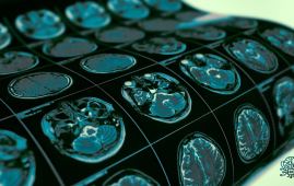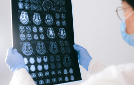

MRI scans provide superior soft tissue contrast to conventional imaging modalities such as X-rays and CT scans. Unfortunately, MRI is extremely sensitive to motion, with even minor movements producing picture distortions. When crucial features are disguised from the clinician, these artifacts put patients at risk of misdiagnosis or incorrect treatment. However, MIT researchers believe they have created a deep learning model capable of motion correction in brain MRI.
“Motion is a common problem in MRI scans,” explains Nalini Singh, an Abdul Latif Jameel Clinic for Machine Learning in Health (Jameel Clinic)-affiliated PhD student in the Harvard-MIT Program in Health Sciences and Technology (HST) and lead author of the paper. “It’s a pretty slow imaging modality.”
MRI scans can range from a few minutes to an hour in length, depending on the type of pictures needed. Small movements can have enormous influence on the resulting image even during the shortest scans. Unlike motion in camera imaging, which usually emerges as a localized blur, motion in MRI frequently results in artifacts that can distort the entire image. Patients may be sedated or asked to limit deep breathing in order to reduce motion. However, these techniques are frequently ineffective in populations that are particularly vulnerable to motion, such as youngsters and individuals with psychiatric illnesses.
“Data Consistent Deep Rigid MRI Motion Correction,” the paper’s title, was just won best oral presentation at the Medical Imaging with Deep Learning conference (MIDL) in Nashville, Tennessee. The method computes a motion-free image from motion-corrupted data without altering the scanning procedure in any way. Singh explains, “Our goal was to combine physics-based modeling and deep learning to get the best of both worlds.”
The significance of this combined approach lies in ensuring consistency between the image output and the actual measurements of what is being depicted; otherwise, the model produces “hallucinations” — images that appear realistic but are physically and spatially inaccurate, potentially worsening diagnostic outcomes.
Obtaining an MRI that is free of motion artifacts, especially from patients suffering from neurological illnesses that generate involuntary movement, such as Alzheimer’s or Parkinson’s disease, would enhance more than simply patient outcomes. According to a research from the University of Washington Department of Radiology, motion affects 15% of brain MRIs. Motion in all types of MRI results in repeated scans or imaging sessions to get images of acceptable quality for diagnosis, which costs the hospital roughly $115,000 per scanner per year.
According to Singh, future research could look into more complex types of head motion as well as motion in other body regions. Fetal MRI, for example, suffers from rapid, unexpected motion that cannot be described using simple translations and rotations.
“This line of work from Singh and company is the next step in MRI motion correction. Not only is it excellent research work, but I believe these methods will be used in all kinds of clinical cases: children and older folks who can’t sit still in the scanner, pathologies which induce motion, studies of moving tissue, even healthy patients will move in the magnet,” says Daniel Moyer, an assistant professor at Vanderbilt University. “In the future, I think that it likely will be standard practice to process images with something directly descended from this research.”
more recommended stories
 Phage Therapy Study Reveals RNA-Based Infection Control
Key Takeaways (Quick Summary) Researchers uncovered.
 Safer Allogeneic Stem Cell Transplants with Treg Therapy
A new preclinical study from the.
 AI in Emergency Medicine and Clinician Decision Accuracy
Emergency teams rely on rapid, accurate.
 Innovative AI Boosts Epilepsy Seizure Prediction by 44%
Transforming Seizure Prediction in Epilepsy Seizure.
 Hypnosis Boosts NIV Tolerance in Respiratory Failure
A New Approach: Hypnosis Improves NIV.
 Bee-Sting Microneedle Patch for Painless Drug Delivery
Microneedle Patch: A Pain-Free Alternative for.
 AI Reshapes Anticoagulation in Atrial Fibrillation Care
Understanding the Challenge of Atrial Fibrillation.
 Hemoglobin as Brain Antioxidant in Neurodegenerative Disease
Uncovering the Brain’s Own Defense Against.
 Global Data Resource for Progressive MS Research (Multiple Sclerosis)
The International Progressive MS Alliance has.
 AI Diabetes Risk Detection: Early T2D Prediction
A new frontier in early diabetes.

Leave a Comment