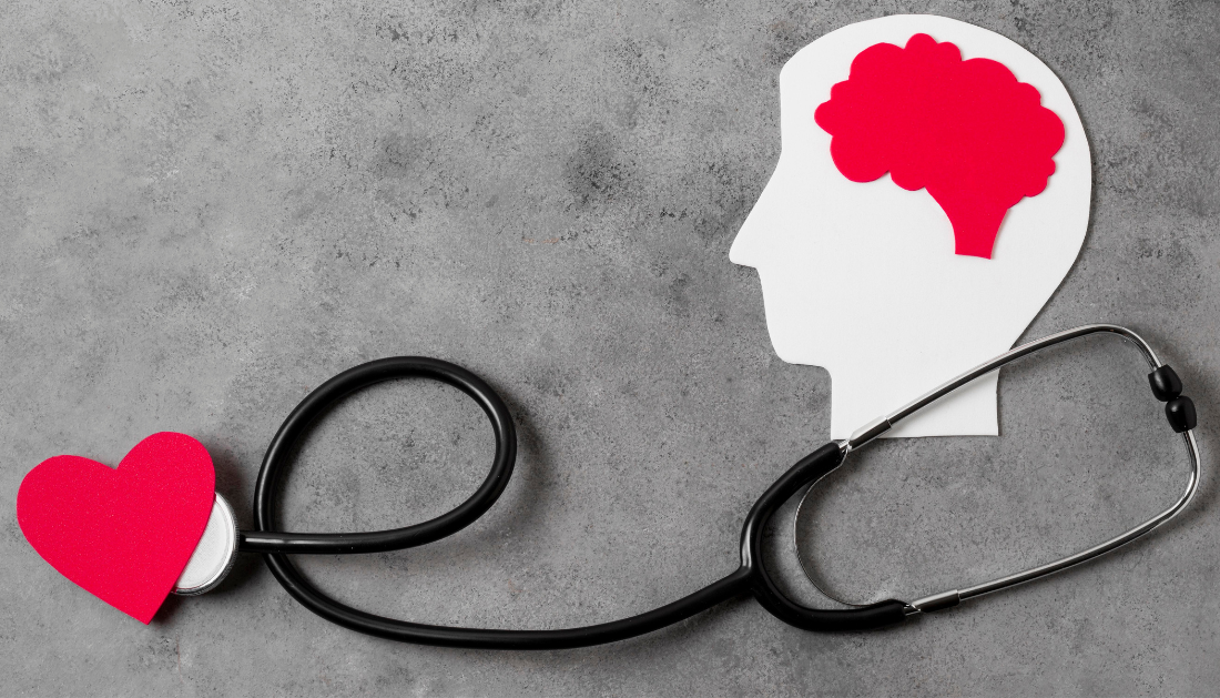

“Our goal was to figure out how to harness TMS treatment more effectively, get the dosing right, by selectively slowing down the heart rate and identifying the individual best spot to stimulate on the brain,” cited senior author Shan Siddiqi, MD, of the Center for Brain Circuit Therapeutics and Brigham’s Department of Psychiatry. According to Siddiqi, the concept came about as a result of Dutch researchers presenting heart-brain coupling results at a conference in Croatia.
“They showed that not only can TMS transiently lower the heart rate, but it matters where you stimulate,” Shan Siddiqi, MD, senior author of the study from the Center for Brain Circuit Therapeutics and the Brigham’s Department of Psychiatry, stated. According to Siddiqi, the concept originated at a symposium in Croatia when academics from the Netherlands were showcasing their heart-brain connection findings.
“They showed that not only can TMS transiently lower the heart rate, but it matters where you stimulate,” The possibility of making this precision-focused treatment for depression more accessible to the rest of the globe is what excites Siddiqi the most about the study, he continued.
“We have so many things we can do with advanced technology available here in Boston to help people with their symptoms,” he said. “But some of those things couldn’t easily get to the rest of the world before.”
“We wanted to see if there would be mostly heart-brain coupling in the connected areas,” Dijkstra said. “For 12 out of 14 usable data sets, we found we would have a very high accuracy of defining an area that is connected by just measuring heart rate during brain stimulation.”
According to Dijkstra, the discovery may aid in the customization of TMS therapy for the treatment of depression by identifying a specific treatment location on the brain and facilitating its accessibility as an MRI would not be required before treatment.
According to Siddiqi, the results of this study could also be utilized in the development of therapies that emergency physicians and cardiologists would find helpful in the future.
The study’s small sample size and the fact that not every region of the brain was stimulated are two of its limitations.
Finding the areas of the brain to activate to improve the consistency of heart rate fluctuations will be the team’s next task.
For more information: Probing prefrontal-sgACC connectivity using TMS-induced heart-brain coupling, Nature Mental Health, https://doi.org/10.1038/s44220-024-00248-8
more recommended stories
 Dietary Melatonin Linked to Depression Risk: New Study
Key Summary Cross-sectional analysis of 8,320.
 Chronic Pain Linked to CGIC Brain Circuit, Study Finds
Key Takeaways University of Colorado Boulder.
 New Insights Into Immune-Driven Heart Failure Progression
Key Highlights (Quick Summary) Progressive Heart.
 Microplastic Exposure and Parkinson’s Disease Risk
Key Takeaways Microplastics and nanoplastics (MPs/NPs).
 Sickle Cell Gene Therapy Access Expands Globally
Key Summary Caring Cross and Boston.
 Reducing Alcohol Consumption Could Lower Cancer Deaths
Key Takeaways (At a Glance) Long-term.
 NeuroBridge AI Tool for Autism Communication Training
Key Takeaways Tufts researchers developed NeuroBridge,.
 Population Genomic Screening for Early Disease Risk
Key Takeaways at a Glance Population.
 Type 2 Diabetes Risk Identified by Blood Metabolites
Key Takeaways (Quick Summary) Researchers identified.
 Microglia Neuroinflammation in Binge Drinking
Key Takeaways (Quick Summary for HCPs).

Leave a Comment