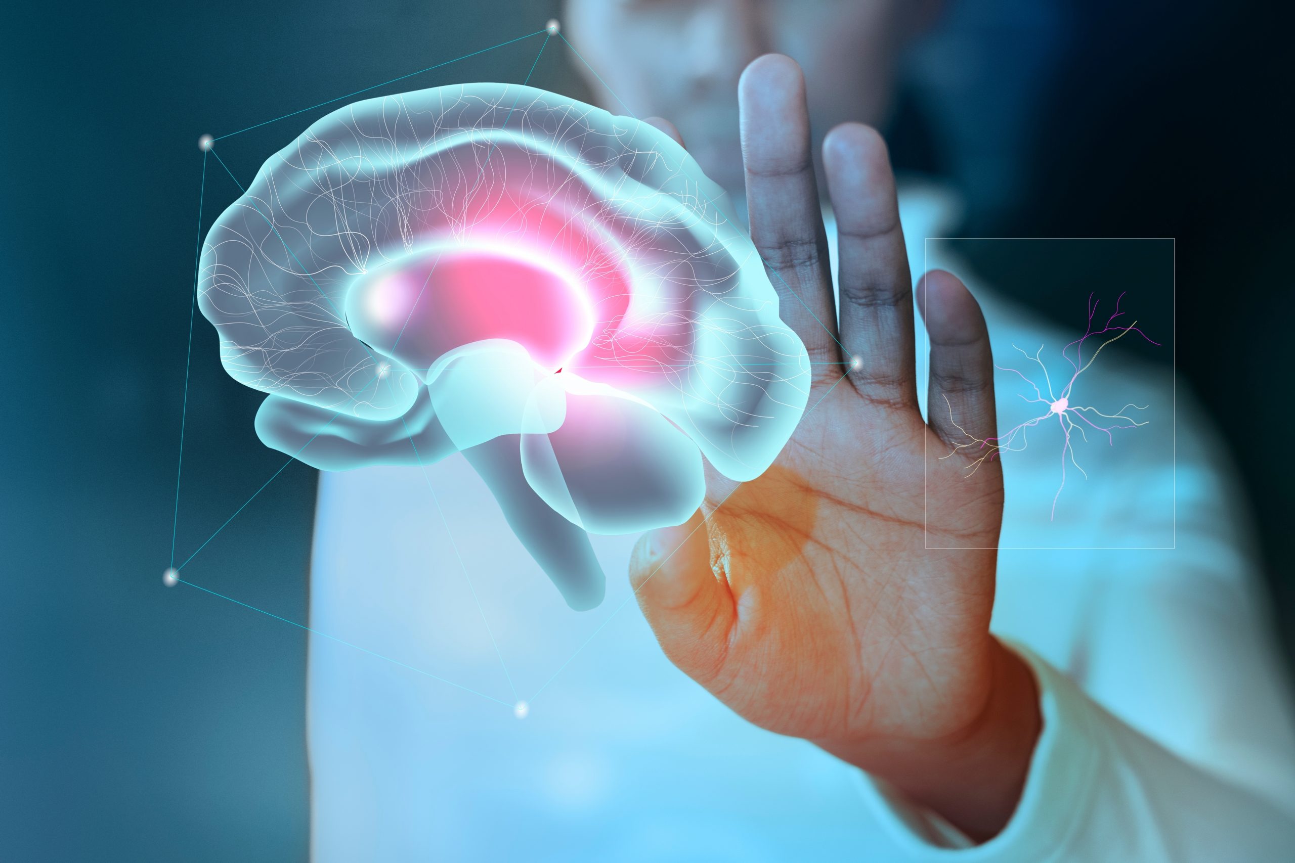
Image by rawpixel on Freepik
The aging mechanisms in the brain have received new insight from researchers at the Trinity Biomedical Sciences Institute (TBSI). They have discovered a potential new target for medicines meant to treat age-related neurological diseases by for the first time connecting the increased presence of specific immune cells to ailments like Alzheimer’s disease and traumatic brain damage.
The study, which concentrated on microglia in the brain and spinal cord and benefited from a partnership with specialists at the University of Maryland School of Medicine, was published today in the journal Science Advances.
The function of microglia, a particular type of immune cell, is to protect against foreign germs, sustain brain cells, remove waste, and engulf and consume dying nerve cells.
New research suggests that the molecular and metabolic alterations taking place inside these specialized cells can cause microglia to respond functionally in various ways.
In actuality, an attribute known as autofluorescence can be used to differentiate different subtypes of microglia. This happens because particular molecules within the cells absorb light, and it describes the propensity of cells to emit light of one color after they have absorbed the light of another. Metals, misfolded proteins, cholesterol crystals, lipid molecules, and other chemicals are all kept in specialized cellular compartments.
The study’s lead author is David Loane, an assistant professor of neuroscience at Trinity’s School of Biochemistry and Immunology (TBSI). He explained, “These materials accumulate inside autofluorescent microglia as the brain ages, which increases their autofluorescence. However, this buildup of cellular debris also makes it more difficult for the microglia to carry out their crucial functions of clearing out waste and preventing neurological damage and neurodegenerative disorders in the brain.
“In this study, we found—in aged animals—that these microglia adopt a unique, dysfunctional state, which has a number of problematic impacts. For example, there is an increase in cellular stress and damage, an accumulation of fats and iron, alterations to metabolic processes, and an increase in production of molecules that over-egg the immune response.”
The researchers also showed that autofluorescent microglia and related inflammation were more evident in pathological settings, such as in genetic risk factor models of neurological diseases like Alzheimer’s disease, and that they could be reversed, in age-related animals, by drug-assisted microglial replacement.
Prof. Loane said, “In addition, environmental exposure to acute traumatic brain injury in animals increased tissue-wide distribution and age of onset of autofluorescent microglia by increasing oxidative stress damage in the damaged animals’ brains.
“As a result, increasing evidence now suggests that the accumulation of autofluorescent microglia contributes to diseases of aging and neurodegeneration. If these sub-populations of microglia are highly inflammatory and damaging to the brain, then targeting them could be a new strategy for treating aging-related diseases.”
 Nanoplastics in Brain Tissue and Neurological Risk
Key Takeaways for HCPs Nanoplastics are.
 AI Predicts Chronic GVHD Risk After Stem Cell Transplant
Key Takeaways A new AI-driven tool,.
 Red Meat Consumption Linked to Higher Diabetes Odds
Key Takeaways Higher intake of total,.
 Pediatric Crohn’s Disease Microbial Signature Identified
Key Points at a Glance NYU.
 Nanovaccine Design Boosts Immune Attack on HPV Tumors
Key Highlights Reconfiguring peptide orientation significantly.
 High-Fat Diets Cause Damage to Metabolic Health
Key Points Takeaways High-fat and ketogenic.
 Acute Ischemic Stroke: New Evidence for Neuroprotection
Key Highlights A Phase III clinical.
 Statins Rarely Cause Side Effects, Large Trials Show
Key Points at a Glance Large.
 Anxiety Reduction and Emotional Support on Social Media
Key Summary Anxiety commonly begins in.
 Liquid Biopsy Measures Epigenetic Instability in Cancer
Key Takeaways Johns Hopkins researchers developed.
Leave a Comment