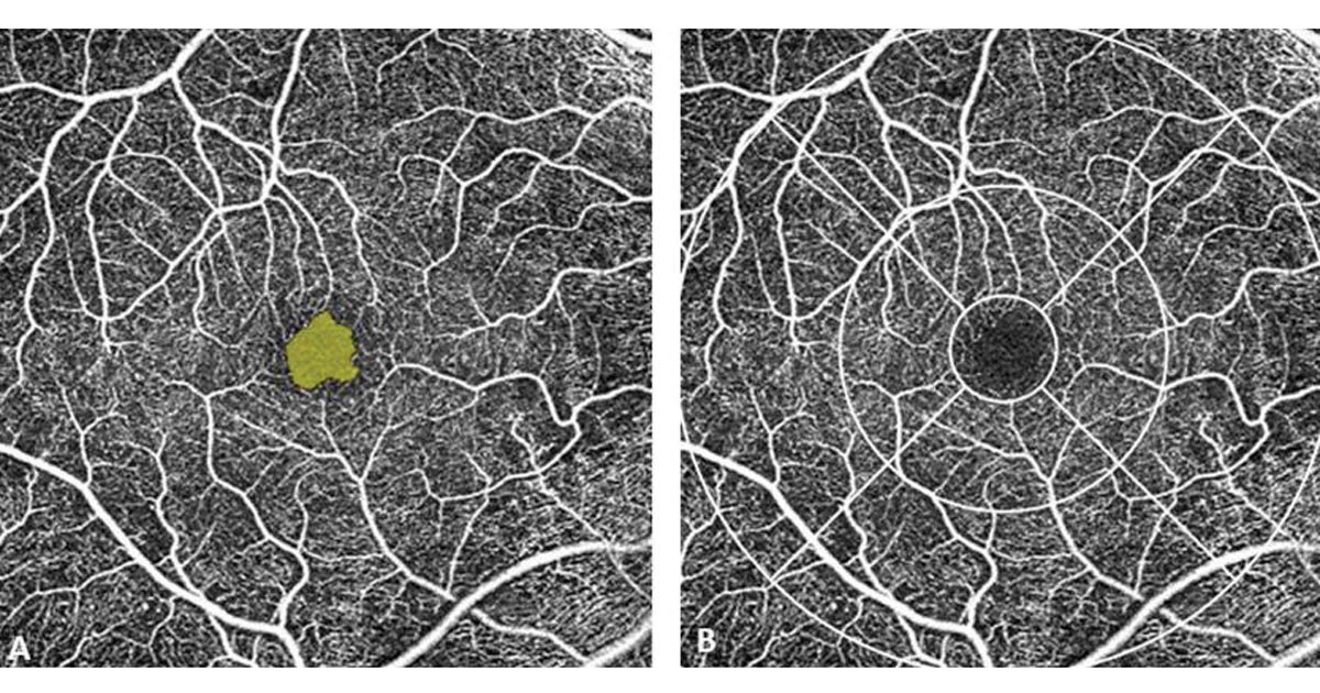

OCTA images showing FAZ boundary drawn using a manual tool (left) and ETDRS grid used to measure vessel density and perfusion (right).
Posterior uveitis is the most common form of involvement in intraocular tuberculosis.
As with all cases of posterior uveitis, imaging plays a key role in documenting lesions, assessing disease activity, determining the location of the disease, detecting complications and sequelae, as well as monitoring response to therapy.
Intraocular inflammation can be associated with various vascular flow abnormalities in a wide spectrum of uveitic disorders. Hence, recognizing patterns of disruption is integral to the diagnosis and management of these conditions. The gold standard modalities for the imaging of retinal and choroidal vessel morphology are fluorescein angiography and indocyanine green angiography. But these procedures are invasive and require the injection of intravenous dyes that may be poorly tolerated and associated with rare serious side effects.
In this column, we highlight the role of OCT angiography (OCTA) in evaluating tuberculous choroiditis.
Tuberculous uveitis
In these cases, tuberculous uveitis had several diagnostic criteria.
1. Ocular findings were consistent with possible intraocular TB with no other cause of uveitis suggested by history, symptoms, or ancillary testing.
2. Diagnosis of TB was made based on one of the following:
- Strong positive tuberculin skin test results (greater than 15-mm area of induration after 48 hours);
- Positive QuantiFERON-TB Gold;
- Microbiological confirmation from sputum or extraocular sites; or
- Chest X-ray consistent with TB infection.
3. In cases of bilateral uveitis, both eyes were included. In unilateral uveitis, only the uveitic eye was included in the analysis.
Exclusion criteria were all known causes of infectious uveitis except TB and known noninfectious uveitis syndrome excluded by clinical features and relevant tests, and patients with poor fixation and media opacities precluding adequate imaging.
A detailed history and clinical examination, including slit-lamp biomicroscopy, fundus examination by 90 D and indirect ophthalmoscopy, IOP and appropriate investigations (ultrasound B-scan, complete blood counts, Mantoux test, erythrocyte sedimentation rate, C-reactive protein, angiotensin-converting enzyme, rheumatoid factor, IgG/IgM toxoplasmosis, venereal disease research laboratory, antinuclear antibodies), were carried out to rule out other causes of uveitis. Standardization of Uveitis Nomenclature Working Group guidelines were used for uveitis anatomical classification and inflammation grading and activity.
OCTA imaging technique
All patients were imaged using AngioPlex (Cirrus HD-OCT 5000, Zeiss). AngioPlex uses the OCT-based microangiography (OMAG) complex algorithm with a central wavelength of 840 nm and an A-scan rate of 68,000 per second. OMAG identifies changes in the phase and intensity information of the OCT scans to quantify motion contrast. For eye-tracking, FastTrac technology was implemented, and the retina was sampled 15 frames per second to minimize motion artefacts. Only areas that may have been affected by motion artefacts were rescanned, which decreased the acquisition time.
The pupil was dilated with a drug combination of tropicamide 0.8% and phenylephrine 5% to target a pupil diameter mean of 6 mm. The A-scan depth was 2 mm with an axial resolution of 5 µm and a transverse resolution of 15 µm. Patients were scanned using 6 mm × 6 mm scans centered on the fovea. The individual vascular layers, namely the superficial capillary plexus (SCP), deep capillary plexus (DCP), choriocapillaris (CC), and choroid, were analyzed using custom segmentation of the device. The representations were SCP at 30 ± 30 µm below the internal limiting membrane (ILM), DCP at 130 ± 12.5 µm below the ILM and retinal pigment epithelium, and CC at 10 ± 0 µm below the basement membrane.
Qualitative image analysis
Segmentation of the SCP, DCP, CC, and choroid layers was inbuilt in the algorithm. These slabs were used to evaluate retinochoroidal blood flow, vasculature, and morphology. The OCTA images were visualized on the AngioPlex software and analyzed to identify distinct morphologic patterns of the retinochoroidal vasculature. The images were analyzed by the same observer to avoid interobserver variations. En face OCT images were compared with structural en face images and cross-sectional OCTA to detect the presence of signal loss and shadowing.
Quantitative analysis
Quantitative analysis of the 6 mm × 6 mm OCT angiograms, including vessel density and perfusion of the superficial retinal plexus (SRP) and foveal avascular zone (FAZ) parameters such as area, perimeter, and circularity, was performed using the AngioPlex Metrix software. In the macular region, vessel density was measured in five sectors within the inner two circles of the ETDRS grid centered on the fovea.
The location of the foveal center was confirmed by cross-referencing with the OCT scans associated with the OCTA image. The vessel density measurements were noted only at the level of the SRP. The area of the FAZ was measured by using the non-flow measurement tool in the software, and in cases in which the FAZ was not detected, a manual drawing tool was used to draw the outline. Central retinal thickness (CRT) was defined as the average thickness of the central 1 mm diameter of the ETDRS grid.

For further analysis, we divided our sample size into two subgroups: anterior uveitis/intermediate uveitis (group A) and posterior uveitis (group B) (Figure 2). Scans with images that had signal intensities of 6/10 or less, large or diffuse floaters, significant segmentation errors, or motion artifacts were excluded.
OCTA vascular observations
The mean CRT was 298.65 ± 160.22 µm. There was no correlation between the vessel density and the CRT in posterior uveitis. However, there was a significant positive correlation with the overall CRT and the vessel density in the center in the superficial plexus. The overall mean vessel density in 48 eyes was 9.4% ± 4.1%. The mean vessel density in posterior uveitis was high compared with the other two groups (anterior and intermediate uveitis). There was statistical significance between the three groups (P = .000). The vessel density central was directly correlated with the vessel perfusion central (P = .000, r = 0.777). Similarly, the inner layer and outer layer vessel density correlated with perfusion (P = .000, r = 0.945). The overall FAZ area was 0.23 ± 0.11 mm2 (Figure 3). The mean foveal perimeter and circularity were 2.6 ± 5.1 µm and 0.63 ± 0.12 µm, respectively. The FAZ area showed negative correlation with the CRT (r = –0.491, P = .001). There was no difference in the FAZ area between the three clinical groups (anterior, intermediate, and posterior uveitis). The mean capillary perfusion density was 22.2% ± 10.2%. There was a significant positive correlation between the capillary perfusion density and the overall superficial vessel density. There was no statistically significant difference in capillary perfusion density between anterior, intermediate, and posterior groups.

Summary
OCTA can be used for the evaluation of vascular changes in eyes with tuberculous choroiditis. OCTA changes can be used for follow-up analysis and prognosis. The noninvasive and noncontact nature of the method makes it more patient-friendly and less complicated. OCTA can present integrated structural and flow information of the human eye in vivo. Besides enabling diagnosis, it also shows great promise in understanding tissue perfusion even in the absence of morphological changes. Thus, OCTA is an important adjunct that supplements the findings of conventional fluorescein angiography and indocyanine green angiography in the diagnosis and management of posterior uveitic entities such as intraocular TB.
more recommended stories
 Texas Medical Board Releases Abortion Training for Physicians
Key Takeaways Texas Medical Board has.
 Safer Allogeneic Stem Cell Transplants with Treg Therapy
A new preclinical study from the.
 Autoimmune Disorders: ADA2 as a Therapeutic Target
Adenosine deaminase 2 (ADA2) has emerged.
 Kaempferol: A Breakthrough in Allergy Management
Kaempferol, a dietary flavonoid found in.
 Early Milk Cereal Drinks May Spur Infant Weight Gain
New research published in Acta Paediatrica.
 TaVNS: A Breakthrough for Chronic Insomnia Treatment
A recent study conducted by the.
 First-of-Its-Kind Gene-Edited Pig Kidney: Towana’s New Life
Surgeons at NYU Langone Health have.
 Just-in-Time Training Improves Success & Patient Safety
A study published in The BMJ.
 ChatGPT Excels in Medical Summaries, Lacks Field-Specific Relevance
In a recent study published in.
 Study finds automated decision minimizes high-risk medicine combinations in ICU patients
A multicenter study coordinated by Amsterdam.

Leave a Comment