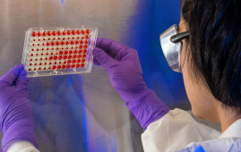

Tulane University School of Medicine researchers have discovered a potential new model for studying fungal pneumonia that has historically been difficult to culture in the lab.
Pneumocystis species, a fungus that causes Pneumocystis pneumonia in immunocompromised patients and children, was studied using precision-cut lung tissue slices.
This breakthrough overcomes a significant barrier in fungal research—the difficulty of growing this pathogen outside of a living lung—allowing scientists to more readily test novel medications to combat the infection. The World Health Organization recently named the fungus one of the top 19 fungal priority infections.
“Pneumocystis is likely the most common fungal pneumonia in children and attempts at culturing the organism have largely not been successful,” said corresponding author Dr. Jay Kolls, John W Deming Endowed Chair in Internal Medicine at Tulane. “Thus, we have not had new antibiotics in over 20 years as they have to be tested in experimental animal studies.”
The Tulane model makes use of precision-cut lung slices that preserve the intricacy and architecture of lung tissue, creating an environment that closely resembles circumstances inside the lung. The findings were reported in the journal mBio.
The troph and ascus forms of the Pneumocystis fungus were grown in mouse tissue for up to 14 days. The viability testing and gene expression research they performed revealed that the fungus survived in the model over time.
“This is the first time both the trophic and ascus forms of Pneumocystis have been maintained long-term outside a mammalian host,” he said.
The model’s suitability for in vitro drug testing was confirmed by the researchers. The expression of Pneumocystis genes was reduced when treated with regularly used drugs trimethoprim-sulfamethoxazole and echinocandins, indicating successful fungus targeting.
The Tulane approach produces several homogeneous lung tissue samples for research from a single lung, allowing for high-capacity testing.
“With optimization, we believe precision lung slices could enable actual growth of Pneumocystis and become a powerful tool for developing new medications to treat this infection,” Kolls said in a statement. “This could significantly accelerate research on this pathogen.”
Ferris T. Munyonho, a Tulane Biomedical Sciences graduate student who received a Fulbright Scholarship after completing his Bachelor of Science from the University of Zimbabwe, led the study.
For more information: Precision-cut lung slices as an ex vivo model to study Pneumocystis murina survival and antimicrobial susceptibility, ASM Journals, https://doi.org/10.1128/mbio.01464-23
more recommended stories
 Nanoplastics in Brain Tissue and Neurological Risk
Key Takeaways for HCPs Nanoplastics are.
 AI Predicts Chronic GVHD Risk After Stem Cell Transplant
Key Takeaways A new AI-driven tool,.
 Red Meat Consumption Linked to Higher Diabetes Odds
Key Takeaways Higher intake of total,.
 Pediatric Crohn’s Disease Microbial Signature Identified
Key Points at a Glance NYU.
 Nanovaccine Design Boosts Immune Attack on HPV Tumors
Key Highlights Reconfiguring peptide orientation significantly.
 High-Fat Diets Cause Damage to Metabolic Health
Key Points Takeaways High-fat and ketogenic.
 Acute Ischemic Stroke: New Evidence for Neuroprotection
Key Highlights A Phase III clinical.
 Statins Rarely Cause Side Effects, Large Trials Show
Key Points at a Glance Large.
 Anxiety Reduction and Emotional Support on Social Media
Key Summary Anxiety commonly begins in.
 Liquid Biopsy Measures Epigenetic Instability in Cancer
Key Takeaways Johns Hopkins researchers developed.

Leave a Comment