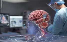
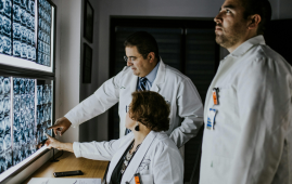
The world’s first sensor prototype that uses gas and laser light to identify flaws in MRI scans is located at Hvidovre Hospital. The novel sensor was created by a young researcher at Hvidovre Hospital and the University of Copenhagen. It can therefore accomplish tasks that are not conceivable for electrical sensors currently in use, which should open the door for more advanced, more affordable, and faster MRI scan.
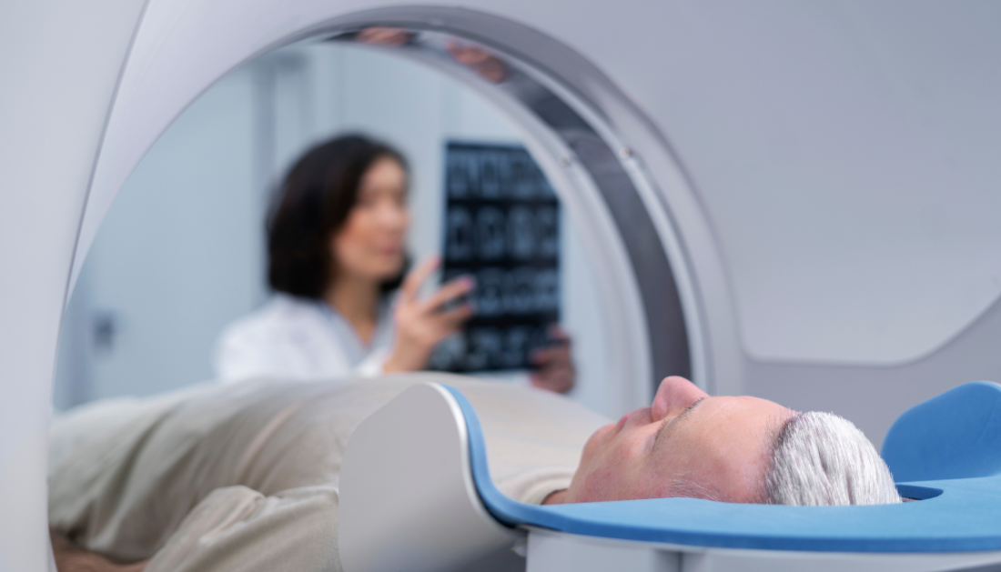
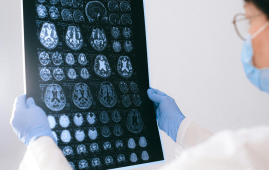
Doctors and other medical professionals utilize MRI scanners on a daily basis to obtain an unparalleled view of the human body. Specifically, they provide 3D images of superior quality compared to other forms of medical imaging, which are utilized to investigate the brain, important organs, and other soft tissues.
There is yet opportunity for improvement, even though this renders the sophisticated instrument priceless and almost vital for medical practitioners.
Variations in the powerful magnetic fields inside MRI scanners lead to mistakes and disruptions in scans. Therefore, in order to minimize errors, these costly machines—which cost hundreds of euros each hour—need to be calibrated on a regular basis.
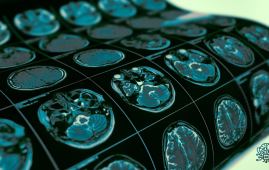
Additionally, there are unique scanning techniques that are regrettably not applicable in modern times. Among these are spiral sequences, which have the potential to shorten scanning times for conditions like tumors, sclerosis, and blood clots. Spiral sequences would also be a desirable tool in MRI research, as they could offer fresh insights into brain disorders to scientists and medical experts, among other uses. However, at this time, it is not possible to execute these kinds of scans because of the extremely unstable magnetic field.
Theoretically, a sensor that maps and reads variations in the magnetic field could fix the issue. After that, using a computer to fix the mistakes in the photographs is really easy. With current technology, this has proven challenging in practice since otherwise appropriate sensors interfere with the magnetic field due to their electric nature and metal cable connections.
This issue is intended to be resolved by a recent invention. A researcher from the Niels Bohr Institute and The Danish Research Centre for Magnetic Resonance (DRCMR) has created a sensor that employs a tiny glass container filled with gas and laser light in fiber cables to fight the issue. The working prototype is ready.
“First we demonstrated that it was theoretically possible, and now we have proven that it can be done in practice. In fact, we now have a prototype that can basically make the measurements needed without disturbing the MRI scanner. It needs to be developed more and fine-tuned, but has the potential to make MRI scans cheaper, better and faster – although not necessarily all three at once,” laughs Hans Stærkind, a postdoc at the Niels Bohr Institute and DRCMR at Hvidovre Hospital. Stærkind is the main architect behind the sensor and device that comes with it.
“An MRI scanner can already produce incredible images if one takes their time. But with the help of my sensor, it is imaginable to use the same amount of time to produce even better imagery – or spend less time and still get the same quality as today. A third scenario could be to build a cheaper scanner that, despite a few errors, could still deliver decent image quality with the help of my sensor,” says the researcher.
How the prototype operates
Protons in the body’s water, proteins, and carbs are forced to align with the magnetic field by the strong magnetic field created by powerful magnets in MRI scanners. Protons are excited when radio waves are pumped through a patient, momentarily spinning them out of equilibrium. They then produce radio waves that can be used to create 3D images of the object being scanned in real time when they align themselves again with the magnetic field.
The working mechanism of Hans Staerkind’s prototype is a laser light transmitter and receiver that resembles a stereo system from the 1990s. It directs laser light into four scanner sensors via fiber optic cables, which are made entirely of glass.
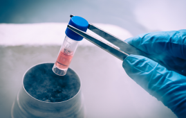
The light enters the sensors through a tiny glass container filled with caesium gas, which absorbs light at the appropriate frequencies.
“When the laser has just the right frequency while passing through the gas, there is a resonance between the waves of light and electrons in the caesium atoms. But the frequency – or wavelength – at which this happens changes when the gas is exposed to a magnetic field. In this way, we can measure the strength of the magnetic field by finding out what the right frequency is. This happens completely automatically and lightning fast by the receiving device,” explains the researcher.
Hans Staerkind’s prototype maps the location of disruptions in the ultra-strong magnetic field of an MRI scanner, as well as the degree to which the field has altered. This may soon imply that erroneous and disrupted images can be rectified using the sensor data, becoming fully functional and precise in the process.
Innovation with business potential—when the necessary data is available
The idea was devised at DRCMR at Hvidovre Hospital in Copenhagen, where the prototype is being kept.
“The original idea came from my supervisor here at DRCMR, Esben Petersen, who is unfortunately no longer with us. He saw huge potential in developing a sensor based on lasers and gas that would be able to measure the magnetic fields without disturbing them,” says Hans Stærkind.
Professor Eugene Polzik and other quantum physicists at the Niels Bohr Institute assisted Staerkind in turning the concept into a working theory. And he has now implemented that theory with the prototype.
“The prototype is designed in such a way that it is already suitable in hospital contexts as a robust and well-functioning instrument. And so far, our tests have shown that it works as it should. One can imagine that this invention will eventually be integrated directly into new MRI scanners,” says Stærkind.
For the time being, more development will be done on the prototype to improve the accuracy of its measurements.
“We need to collect data and fine-tune it so that it continuously becomes a better and better tool for finding errors in scans. After that, we’ll move on to the exciting work of correcting errors in MRI images, and find out in what situations and which types of scans our sensor can make a significant difference,” says the researcher.
MRI research units are Staerkind’s primary target market for his sensor. In the slightly longer future, though, he also hopes that one of the major MRI producers finds out about the new technology.
“Once the prototype has been refined in a 2.0 version and its qualities documented with plenty of data from actual scans here at the hospital, we will see where this goes. It certainly has the potential to improve MRI scans in a unique way that can benefit doctors and, not least, patients,” says the researcher.
more recommended stories
 Phage Therapy Study Reveals RNA-Based Infection Control
Key Takeaways (Quick Summary) Researchers uncovered.
 Safer Allogeneic Stem Cell Transplants with Treg Therapy
A new preclinical study from the.
 AI in Emergency Medicine and Clinician Decision Accuracy
Emergency teams rely on rapid, accurate.
 Innovative AI Boosts Epilepsy Seizure Prediction by 44%
Transforming Seizure Prediction in Epilepsy Seizure.
 Hypnosis Boosts NIV Tolerance in Respiratory Failure
A New Approach: Hypnosis Improves NIV.
 Bee-Sting Microneedle Patch for Painless Drug Delivery
Microneedle Patch: A Pain-Free Alternative for.
 AI Reshapes Anticoagulation in Atrial Fibrillation Care
Understanding the Challenge of Atrial Fibrillation.
 Hemoglobin as Brain Antioxidant in Neurodegenerative Disease
Uncovering the Brain’s Own Defense Against.
 Global Data Resource for Progressive MS Research (Multiple Sclerosis)
The International Progressive MS Alliance has.
 AI Diabetes Risk Detection: Early T2D Prediction
A new frontier in early diabetes.
Leave a Comment