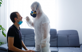
Image by kjpargeter on Freepik
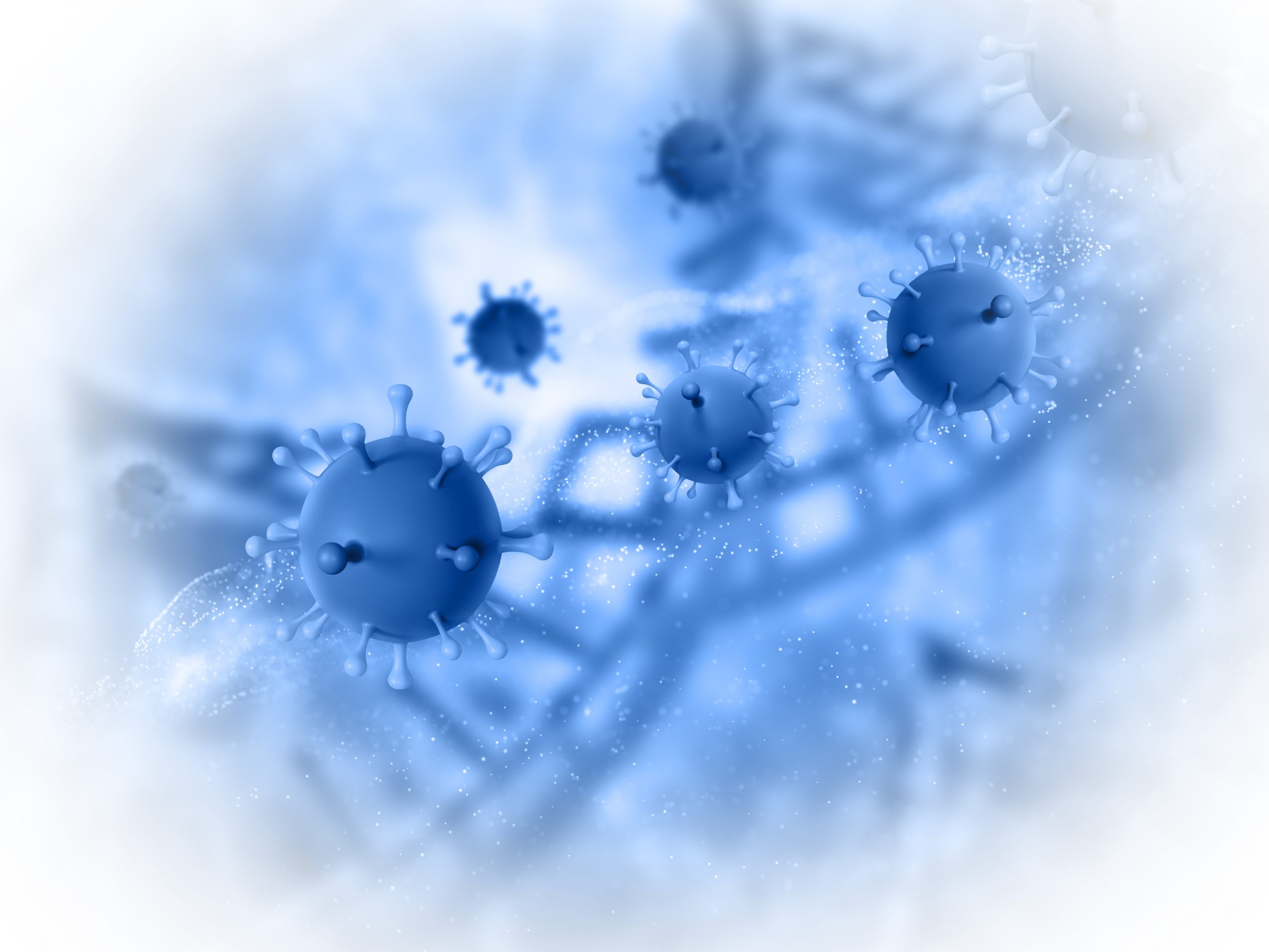
The SARS-CoV-2 virus works really hard to replicate as soon as it infects a cell in the throat or lungs. It uses the metabolic pathways of the host cell to manufacture its proteins and ensure that its genetic material (the RNA genome) is replicated. After that, the newly formed virus particles are ejected from the cell to infect further cells with their highly compressed RNA genome.
The nucleocapsid protein (N), a specific viral protein, is crucial for quick and effective replication. It ensures that the virus’s extremely long RNA is tightly coiled by wrapping around the RNA genome of the organism. When N enters the cell, it separates from the RNA genome and takes on a variety of roles during viral replication.
N shields the RNA from being damaged by the cell’s antiviral defense system (“RNA interference”) when the RNA transforms into viral proteins. Finally, N gathers the SARS-CoV-2 virus that has been replicated in the cell and coils it allowing the formation of new viral particles. N also directly contributes to RNA transcription into viral proteins.
N can carry out all of these functions with a variety of tools at its disposal, similar to a Swiss army knife: First, N needs to be able to tell the difference between viral and cellular RNA and coil the latter into a spiral shape. This explains why N can bind the SARS-CoV-2 virus RNA in a rather indiscriminate manner. For instance, N must, however, equally control the translation of viral RNA into viral proteins.
The N-terminal domain (NTD), one of N’s tools, has been used by researchers led by Dr. Sophie Korn and Andreas Schlundt from the Institute for Molecular Biosciences and the Center for Biomolecular Magnetic Resonance (BMRZ) at Goethe University Frankfurt to shed light on how this particular binding operates. Their findings add to earlier research by the COVID-19-NMR group, founded in Frankfurt amid the epidemic.
The atoms of the NTD tool and the bound RNA are exposed to an intense magnetic field in the work by Korn and her colleagues, who then used nuclear magnetic resonance (NMR) spectroscopy, which reveals information about their spatial arrangement during binding.
This work was recently published in Nature Communications.
Small-angle X-ray scattering, or SAXS, a specialized X-ray technique, also provided detailed data regarding the stability of the newly created molecular complexes.
The end result: The positively charged portion of the NTD contacts the negatively charged RNA in a very generic manner since the RNA building blocks (bases) sequence and the spatial folding of the RNA are critical for binding. The NTD then probes the RNA using a number of “fingers” to look for patterns that will help it bind to the RNA more firmly.
The NTD favors motifs in lung cells at body temperature in a particular spatial folding that is lost when the temperature rises by just a few degrees, which caught the attention of the researchers. This makes them more closely bound and designates them as their own target motifs, which may allow the NTD to do new tasks.
Sophie Korn says, “Although our data are only a first step, they suggest that the virus could switch between replication and packaging into new virus particles in this way: At normal body temperature, the cell predominantly produces building blocks for new viruses.
“If we develop a fever in the course of the infection because our immune system recognizes and fights the virus, the virus might switch to replication as a direct result and ensure that the viral RNA is packaged more and released in the form of new virus particles. It is the viral RNA motifs themselves that provide the switch, which is then triggered by the human defense system.”
Andreas Schlundt states, “With the combination of NMR spectroscopy and SAXS, we have established an experimental method that we can use to quickly assess which binding partners N prefers, and this can probably be transposed to other viral proteins. This will be useful both in the study of emerging viruses and viral variants as well as in the development of antiviral drugs that systematically disable the virus, minimizing side effects in the process.”
 Study Reveals Cold May Impact SARS-CoV-2 Infection Rates
The Unexpected Protective Role of Rhinoviruses.
 Heart, Lung, & Brain Risks Persist in COVID-19 Survivors
A French nationwide study reveals that.
 How COVID-19 and Vaccines Differ in Heart Inflammation
A team of international researchers led.
 Long COVID: Extended Paxlovid Treatment Offers Hope
A new case series by UC.
 RSV Vaccine Response in Immunocompromised Adults
According to Johns Hopkins Medicine researchers,.
 Gut Microbiome Predicts Long COVID Risk
In a recent pre-print study published.
 COVID-19 & Autoimmune Care Hope: Natural Proteins
Recent research at Umeå University reveals.
 FasL Inhibitor Asunercept Speeds COVID-19 Recovery
A new clinical trial demonstrates that.
 Impact of COVID-19 mRNA Vaccine on Myocardial Scarring
A new study found a greater.
 Long-term Cognitive and Psychiatric Issues in COVID-19 Survivors
A new study published in The.
Leave a Comment