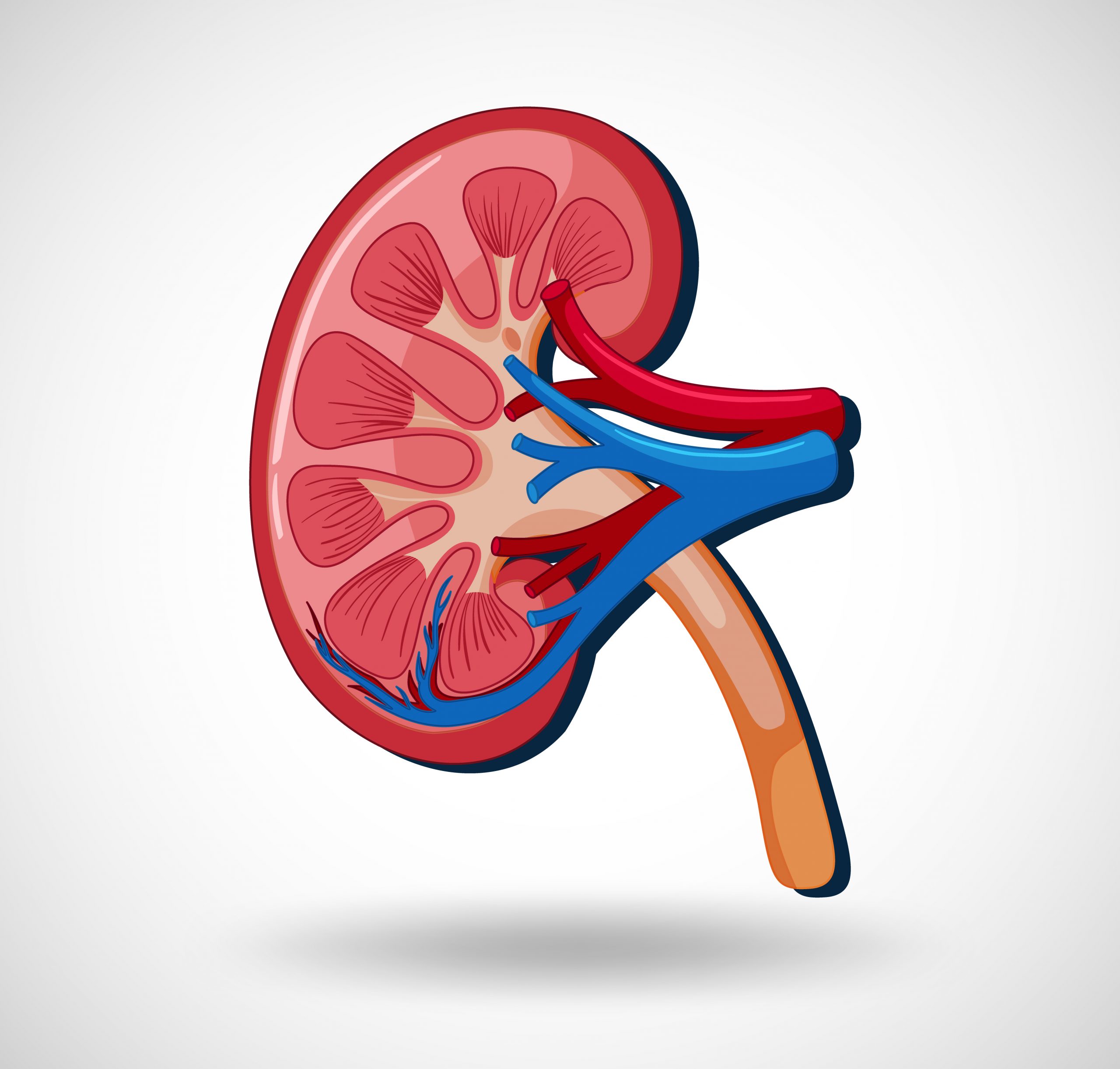

Scientists at Washington University School of Medicine in St. Louis are among the leaders of a multi-institutional research team that has created an atlas of the kidney’s countless cells. The goal of the kidney tissue atlas is to improve understanding of kidney injury and disease.
Building on prior research that identified 30 cell kinds in the kidney, the researchers discovered 51 cell types, some rare and novel, in the healthy kidney and 28 related cell types with traits associated with injury or recovery. They discovered various cellular microcosms with rich genetic signatures along parts of the kidney related with renal failure in patients or recovery from injury. They generated a complete 2D and 3D atlas of kidney cell arrangement and molecular identification in healthy and sick kidneys.
The study, funded by the National Institute of Diabetes and Digestive and Kidney Diseases of the National Institutes of Health (NIH), was published in Nature on July 19th.
“We don’t have great treatment options for patients with kidney disease,” said Sanjay Jain, MD, PhD, a Washington University professor of medicine who led this study with five co-corresponding authors. “By mapping molecular signatures, we hope to predict which patients are at risk of progressing to kidney failure. This molecular knowledge will, one day, lead us to precise, customized treatments for our patients.”
Jain’s team, which included co-investigators Joseph P. Gaut, MD, PhD, the Ladenson Professor of Pathology & Immunology; Anitha Vijayan, MD, a professor of medicine in the Division of Nephrology; and Eric H. Kim, MD, an associate professor of surgery, collaborated with members of the Kidney Precision Medicine Project (KPMP), Human BioMolecular Atlas Program (HuBMAP), Human Cell Atlas, and scientists from other institutions to perform single-cell
The NIH-funded Kidney Precision Medicine Project attempts to improve kidney disease treatment. It is attempting to accomplish this with the assistance of kidney illness participants who are willing to undergo kidney biopsies only for research purposes. HuBMAP, an NIH Common Fund-sponsored program that aspires to spatially identify every cell in a healthy human body, and the Human Cell Atlas, an international scientific endeavor to collect information on at least 10 billion human cells, both provided funding for the work.
The new study is one of nine articles published simultaneously across Nature Portfolio journals that introduce the first set of maps developed by researchers from HuBMAP-supported institutions. Jain is also a co-senior author on another study, published in Nature Communications, that maps healthy and injured cellular communities in kidney stone formation sites and discovers biomarkers in urine that are specific to patients with kidney stone disease.
After the age of 35, kidney function gradually falls. Chronic illnesses like diabetes and hypertension, as well as accumulating kidney injury from physical impact, low oxygen or changes in blood flow to the kidneys during surgery, medication reactions, recreational drug use, and even dehydration, can rapidly accelerate natural decline and lead to kidney disease and, eventually, kidney failure. Dialysis or difficult-to-obtain organ donation are the only treatment alternatives.
The researchers used single-cell studies to identify the molecular characteristics of healthy and sick kidney cells in various kidney segments from patient kidney biopsy samples. They also used spatial analysis to generate 3D images of cells living in communities and communicating with one another. A full understanding of such interactions could help to reduce nonspecific therapeutic targeting of the entire kidney and cellular populations, paving the path for better, more targeted medications with fewer adverse effects.
“We created a benchmark atlas that the community can use as a reference to get insights into how kidney disease develops,” said Jain, who also serves as the director of the Kidney Translational Research Center, which serves as a biorepository for collected kidney biopsies, some of which were used in the study as part of the Human BioMolecular Atlas Program. “We investigated the organization of kidney cells, their molecular identities, and how they transition from healthy to diseased states.” With this information, we can begin to consider medications or small molecule targets that can prevent disease development or improve kidney injury recovery.”
Jain was astounded by the variety of cell types discovered by studying 300,000 human kidney cells, including interstitial, immunological, and endothelial cells. “We found several altered cell states in many different kidney segments, reflecting the plasticity in cell phenotypes as cells transition through healthy, kidney injury, and recovery states,” he said, adding that he is amazed by the complexity of the atlas’ second version, which is currently in the works and involves analyzing more than 1 million kidney cells.
The partnership has resulted in the development of many resources that users can use to identify and comprehend their own study findings. One such program offers consistent nomenclature and benchmark identities for major, minor, and rare kidney cells. Researchers can now annotate their own data sets, assisting in the promotion of uniform communication among research studies.
more recommended stories
 Silica Nanomatrix Boosts Dendritic Cell Cancer Therapy
Key Points Summary Researchers developed a.
 Vagus Nerve and Cardiac Aging: New Heart Study
Key Takeaways for Healthcare Professionals Preserving.
 Cognitive Distraction From Conversation While Driving
Key Takeaways (Quick Summary) Talking, not.
 Fat-Regulating Enzyme Offers New Target for Obesity
Key Highlights (Quick Summary) Researchers identified.
 Spatial Computing Explains How Brain Organizes Cognition
Key Takeaways (Quick Summary) MIT researchers.
 Gestational Diabetes Risk Identified by Blood Metabolites
Key Takeaways (Quick Summary for Clinicians).
 Phage Therapy Study Reveals RNA-Based Infection Control
Key Takeaways (Quick Summary) Researchers uncovered.
 Pelvic Floor Disorders: Treatable Yet Often Ignored
Key Takeaways (Quick Summary) Pelvic floor.
 Urine-Based microRNA Aging Clock Predicts Biological Age
Key Takeaways (Quick Summary) Researchers developed.
 Circadian Control of Neutrophils in Myocardial Infarction
Key Takeaways for HCPs Neutrophil activity.

Leave a Comment