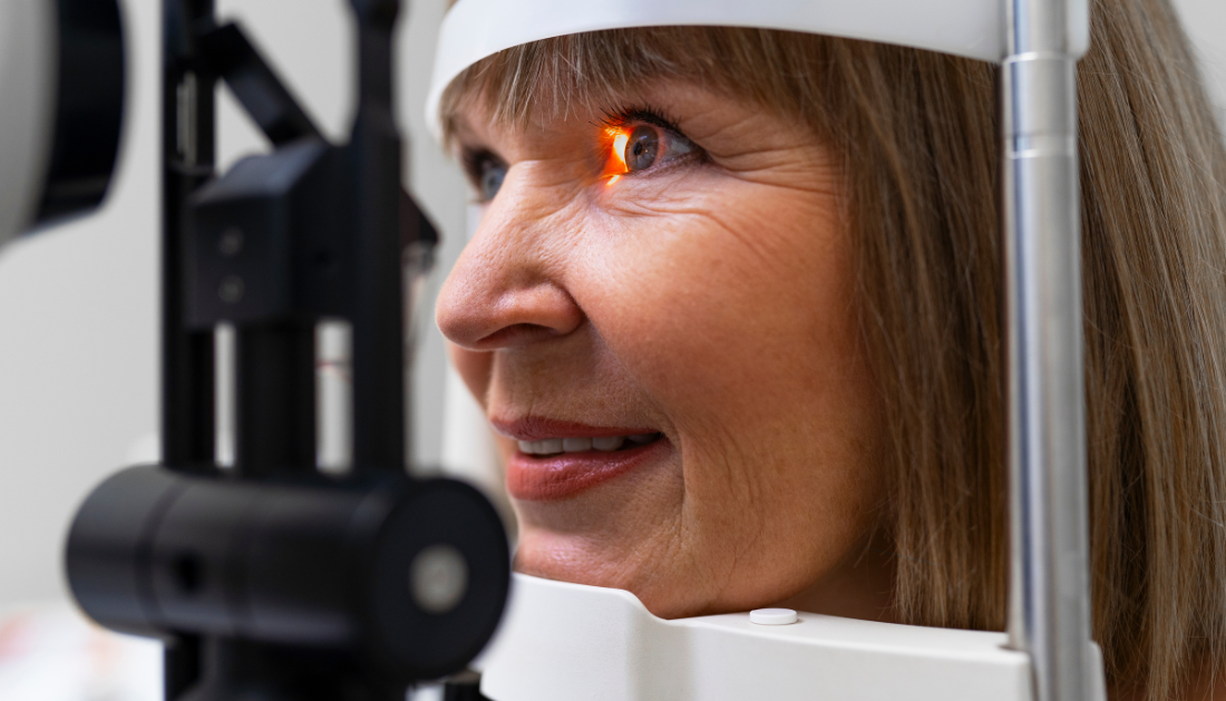

For the first time, research conducted by the University Hospital Bonn (UKB) and the University of Bonn has demonstrated that specific early alterations in individuals suffering from age-related macular degeneration (AMD) might result in a quantifiable local loss of vision. This finding may aid in the testing of novel treatments as well as the management and observation of this eye condition in elderly people, which would otherwise cause central blindness gradually.
AMD primarily affects the elderly. The illness causes a progressive loss of central vision if left untreated, which makes it extremely difficult to do daily tasks like reading or driving. Researchers from all across the world are working hard to find ways to enhance the disease’s early diagnosis and treatment before significant losses happen.
Patients with early forms of AMD have been carefully studied by a UKB Eye Clinic research team in close collaboration with basic and clinical experts, as well as with the University of Bonn. The so-called iRORA lesions, which are extremely early anatomical indicators of retinal degeneration, were the subject of the study.
The examinations were performed by Drs. Leon von der Emde, Marlene Saßmannshausen, and Julius Ameln. “We used the microperimetry method to precisely measure the visual acuity at these affected areas of the retina,” the doctors add. To detect visual abnormalities, this includes assessing the retina’s sensitivity to light stimuli. Routine clinical equipment reach their limits when the damaged retinal regions are less than 250 micrometers.
Assistance is provided via an adaptive optics scanning light ophthalmoscope (AOSLO), a high-resolution research tool created in Bonn. “It allows functional testing of small areas down to individual photoreceptors and allows microscopic resolution imaging of the retina,” says Dr. Wolf Harmening, head of the AOSLO laboratory at the UKB Eye Hospital and a member of the University of Bonn’s Transdisciplinary Research Area (TRA) “Life & Health.”
The outcome was evident: there was a noticeable decrease in visual acuity in the lesioned areas. In comparison to a control region, the loss using the traditional method was, on average, 7 units. The loss using the exact AOSLO approach was 20, which is equivalent to a factor of 100 decrease in light sensitivity.
These findings demonstrate that eyesight is already significantly affected by iRORA lesions. Early retinal damage may act as a marker to help track the disease’s progression and initiate treatment early on. The findings of this investigation represent a significant advancement in our comprehension of the mechanisms behind the development of substantial retinal damage in the late type of dry AMD.
“Our investigations show that even these early lesions can contribute to a very localized but nonetheless significant deterioration in vision in our patients.”- Dr. Wolf Harmening, University of Bonn
“This makes them a potential marker that can help to better monitor the progression of AMD and treat it at an earlier stage,” adds Prof. Dr. Frank Holz, Director of the UKB Eye Clinic.
For more information: Assessment of local sensitivity in incomplete retinal pigment epithelium and outer retinal atrophy (iRORA) lesions in intermediate age-related macular degeneration (iAMD), BMJ Open Ophthalmology, https://doi.org/10.1136/bmjophth-2024-001638
more recommended stories
 Red Blood Cells Improve Glucose Tolerance Under Hypoxia
Key Takeaways for Clinicians Chronic hypoxia.
 Nanoplastics in Brain Tissue and Neurological Risk
Key Takeaways for HCPs Nanoplastics are.
 AI Predicts Chronic GVHD Risk After Stem Cell Transplant
Key Takeaways A new AI-driven tool,.
 Red Meat Consumption Linked to Higher Diabetes Odds
Key Takeaways Higher intake of total,.
 Pediatric Crohn’s Disease Microbial Signature Identified
Key Points at a Glance NYU.
 Nanovaccine Design Boosts Immune Attack on HPV Tumors
Key Highlights Reconfiguring peptide orientation significantly.
 High-Fat Diets Cause Damage to Metabolic Health
Key Points Takeaways High-fat and ketogenic.
 Acute Ischemic Stroke: New Evidence for Neuroprotection
Key Highlights A Phase III clinical.
 Statins Rarely Cause Side Effects, Large Trials Show
Key Points at a Glance Large.
 Anxiety Reduction and Emotional Support on Social Media
Key Summary Anxiety commonly begins in.

Leave a Comment