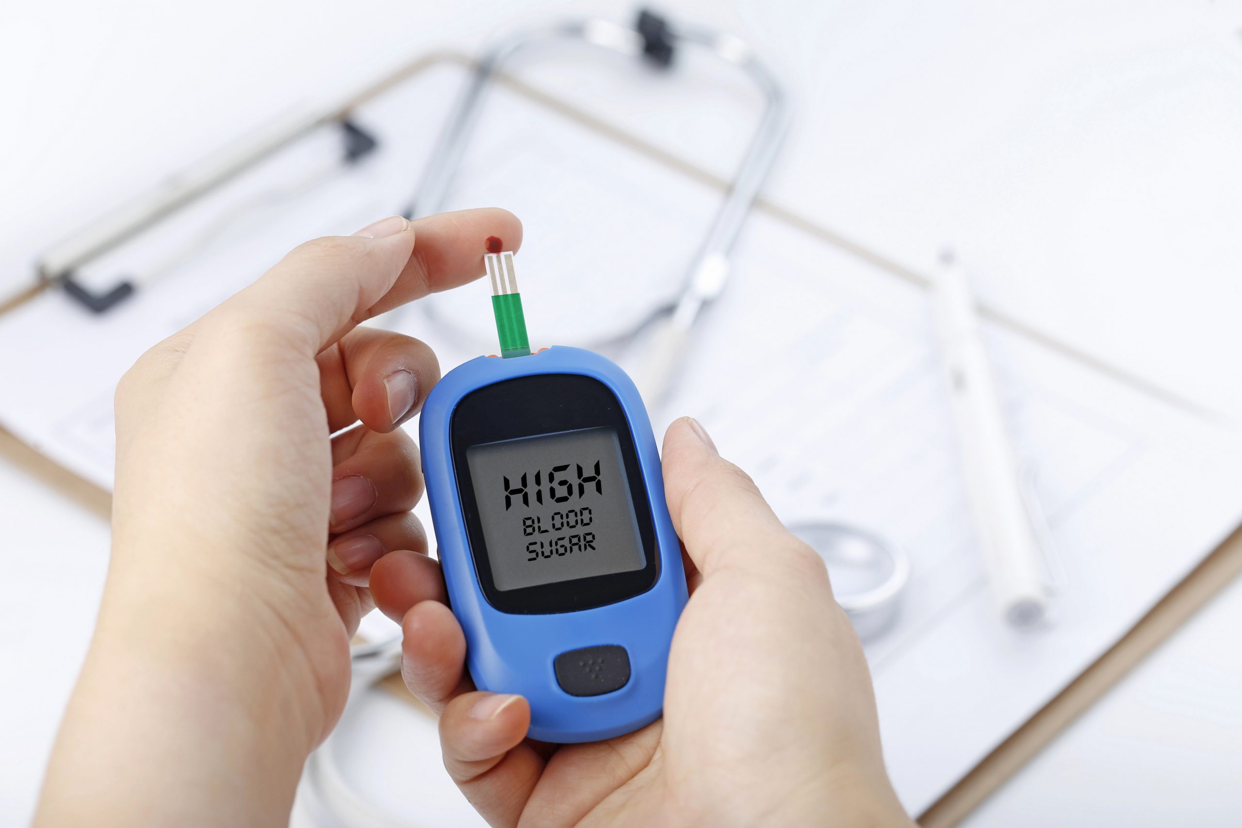

According to a new artificial intelligence model, even in people who don’t fit the criteria for heightened risk, X-ray pictures taken as part of routine medical care can reveal diabetes warning symptoms. According to a multi-institutional team that published their findings in Nature Communications, the model could aid doctors in identifying diabetes illness early and avoiding complications.
The researchers created a model that successfully indicated higher diabetes risk in a retrospective analysis, frequently years before patients were actually diagnosed with the disease. They did this by using the computational technique known as deep learning to photographs and electronic health record data. Given that the prevalence of diabetes in the U.S. has more than doubled over the past 35 years, the researchers think that is noteworthy.
According to current recommendations, people should be screened for type 2 diabetes if they are between the ages of 35 and 70 and have a body mass index (BMI) between the range of overweight and obese.
However, numerous studies have indicated that this approach misses a large proportion of cases, especially among racial/ethnic minorities for whom BMI is a less reliable predictor of diabetes risk. Patients who have untreated diabetes are far more likely to experience fatal consequences, including irreparable organ damage.
Millions of Americans have chest X-rays every year for injuries, breathing problems, chest pain, and in preparation for surgery. Every year, Emory alone completes around 200,000 radiographs. Radiologists examine these X-rays without specifically looking for diabetes, but the images form a part of the patient’s medical file and may be checked later for diabetes or other problems.
“Chest X-rays provide an ‘opportunistic’ alternative to universal diabetes testing,” says Judy Wawira Gichoya, MD, assistant professor of radiology and imaging sciences, and the lead researcher from Emory. “This is an exciting potential application of AI to pull out data from tests used for other reasons and positively impact patient care.”
More than 270,000 X-ray pictures from 160,000 patients were used to train the AI model, and deep learning was used to identify the image elements that most accurately predicted a diabetes diagnosis in the future. The researchers also employed explainable AI techniques to ascertain how and why the model arrived at its conclusions because chest X-rays are not frequently utilized to detect diabetes.
A recent medical finding that visceral fat in the upper body and belly is linked to type 2 diabetes, insulin resistance, hypertension, and other illnesses is consistent with the techniques’ assertion that the location of fatty tissue is crucial for evaluating risk.
The researchers who first created the model resorted to the Emory team to do external validation of the model because of the novelty of the technique and the unexpected outcomes. The Emory researchers discovered that the model predicted risk better than a straightforward model based solely on non-image clinical data when it was applied to a second set of almost 10,000 patients.
In other instances, the chest X-ray forewarned of increased diabetes risk up to three years before to the patient’s eventual diagnosis. The model’s output also offers a numerical risk score that may enable clinicians to adapt the patient’s treatment strategy.
“Diabetes is a chronic disease where body fat distribution matters. The longer the duration of the disease and the worse the glycemic control, the higher the risk of complications,” says Francisco Pasquel, MD, associate professor in Emory’s Division of Endocrinology, Metabolism and Lipids. “The opportunistic approach of using chest X-rays to identify those at the highest risk of diabetes, even before a spike or drop in blood sugar levels occurs, is a promising method that may help improve outcomes through early preventive measures or treatment.”
In order to inform physicians to pursue traditional diabetes screening of individuals labeled as high risk based on X-ray results, the study team will now look at how to further validate the model and incorporate it into electronic health record systems.
After that, they’ll look into how well chest X-rays can be used to identify other diseases like vascular disease, congestive heart failure, and chronic obstructive pulmonary disease.
more recommended stories
 Statins Rarely Cause Side Effects, Large Trials Show
Key Points at a Glance Large.
 Can Too Many Antioxidants Harm Future Offspring?
Key Takeaways High-dose antioxidant supplementation in.
 Anxiety Reduction and Emotional Support on Social Media
Key Summary Anxiety commonly begins in.
 Liquid Biopsy Measures Epigenetic Instability in Cancer
Key Takeaways Johns Hopkins researchers developed.
 Human Antibody Drug Response Prediction Gets an Upgrade
Key Takeaways A new humanized antibody.
 Pancreatic Cancer Research: Triple-Drug Therapy Success
Key Summary Spanish researchers report complete.
 Immune Cell Epigenome Links Genetics and Life Experience
Key Takeaway Summary Immune cell responses.
 Dietary Melatonin Linked to Depression Risk: New Study
Key Summary Cross-sectional analysis of 8,320.
 Chronic Pain Linked to CGIC Brain Circuit, Study Finds
Key Takeaways University of Colorado Boulder.
 New Insights Into Immune-Driven Heart Failure Progression
Key Highlights (Quick Summary) Progressive Heart.

Leave a Comment