
Study: An in vitro characterization of a PCL-fibrin scaffold for myocardial repair
Scientists at the University of Colorado Anschutz Medical Campus have developed a full-thickness, biodegradable patch that has the potential to treat congenital heart defect in neonates while reducing invasive operations and outlasting current patches.
The findings were published in the journal Materials Today this week.
The ultimate goal is to make lab-grown heart tissue from a patient’s own cells that can be used to restructure the heart to correct for heart defects.”
Jeffrey Jacot, PhD, study’s senior author, associate professor of bioengineering, University of Colorado School of Medicine
“The current patch materials available to pediatric heart surgeons are exclusively non-living and non-degradable, which often fail in their long-term therapeutic efficacy due to low compliance, an increased risk of thrombosis and intimal hyperplasia, and their inability to remodel and integrate with the heart,” the study said.
Permanent repairs necessitate biomaterials that are biodegradable but also encourage heart regeneration, so that the patches are eventually replaced by healthy myocardium, the heart’s middle and thickest muscular layer.
“Any patches that are not replaced by healthy tissue prior to their degradation will inevitably fail and lead to long-term complications,” Jacot said.
The patch was generated in the lab using an electrospinning technology, in which electricity is used to liquid solutions to create nanofibers that are then utilized to make a’scaffold.’ Living cells are then inserted into the scaffold. This is finally turned into the patch.
“The scaffold was found to be mechanically sufficient for heart wall repair,” Jacot said. “Vascular cells were able to infiltrate more than halfway through the scaffold in static culture within three weeks.”
More testing is required before the patch may be utilized in humans.
Jacot believes it will play an important role in the treatment of congenital heart abnormalities and other cardiac disorders in the future.
“This is the first successful demonstration of a very thick, porous electrospun patch specifically for cardiac tissue engineering,” he said.
Jarrell, D. K., & Jacot, J. G. (2023). An in vitro characterization of a PCL-fibrin scaffold for myocardial repair. Materials Today Communications. doi.org/10.1016/j.mtcomm.2023.107596.
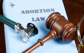 Texas Medical Board Releases Abortion Training for Physicians
Key Takeaways Texas Medical Board has.
 Phage Therapy Study Reveals RNA-Based Infection Control
Key Takeaways (Quick Summary) Researchers uncovered.
 Safer Allogeneic Stem Cell Transplants with Treg Therapy
A new preclinical study from the.
 AI in Emergency Medicine and Clinician Decision Accuracy
Emergency teams rely on rapid, accurate.
 Innovative AI Boosts Epilepsy Seizure Prediction by 44%
Transforming Seizure Prediction in Epilepsy Seizure.
 Hypnosis Boosts NIV Tolerance in Respiratory Failure
A New Approach: Hypnosis Improves NIV.
 Bee-Sting Microneedle Patch for Painless Drug Delivery
Microneedle Patch: A Pain-Free Alternative for.
 AI Reshapes Anticoagulation in Atrial Fibrillation Care
Understanding the Challenge of Atrial Fibrillation.
 Hemoglobin as Brain Antioxidant in Neurodegenerative Disease
Uncovering the Brain’s Own Defense Against.
 Global Data Resource for Progressive MS Research (Multiple Sclerosis)
The International Progressive MS Alliance has.
Leave a Comment