Study: Primate superior colliculus is causally engaged in abstract higher-order cognition
They were shocked to discover that the SC was even more active than the PPC in influencing the respondents’ choices of categories, indicating that it may assist in coordinating higher-order cognitive functions that are generally believed to occur in the neocortex.
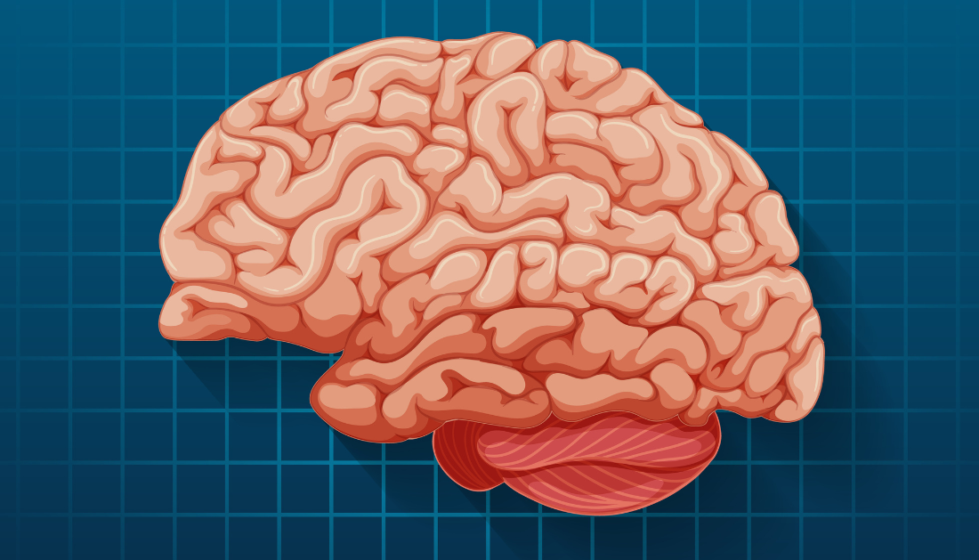
“This is a really surprising place to find these kinds of cognitive signals because this area of the brain is traditionally associated with simpler spatial orienting behaviors and even reflexive functions,” said David Freedman, Ph.D., Professor of Neurobiology and the Neuroscience Institute at UChicago and senior author of the new study.
“We have this evolutionarily ancient brain structure that seems to be even more involved in complex cognitive decisions than the cortical areas we studied in our experiments.”
An old brain area possessing unexpected abilities
Every species, including humans, fish, and reptiles, as well as mammals like primates, must be able to rapidly identify and classify items in their field of vision. Is what is approaching them a threat or an impediment? What’s that darting by, prey or predator?
All vertebrates, including those lacking more complex neocortex, have evolved to have a part of the brain called the SC. In addition to aiding in the orientation of head and eye movements toward visual cues, it was formerly thought to initiate reflexive motor responses by transmitting signals from brain regions upstream.
Recent studies have revealed that it is also involved in more difficult activities, such as choosing a point of orientation and focusing on stimuli in various geographical locations.
For many years, Freedman and his colleagues have been researching other cortical regions that have close anatomical ties to the SC. The researchers aimed to determine whether the SC is also involved in more abstract thinking, since these nearby regions are involved in flexible and cognitively demanding decision-making tasks.
In the most recent study, they trained primates to examine images on a computer screen and make visual decisions. After pressing a button at the appropriate moments to classify photos into the appropriate categories, the animals were rewarded with fruit juice.
The SC and the lateral intraparietal area (LIP), a region of the PPC that Freedman’s group has previously demonstrated is involved in category decisions during these kinds of activities, were the sites of brain cell activity that the researchers saw as the subjects completed the experiment.
The experimental design focused on the brain activity necessary for classification rather than the eye or head movements that are typically assumed to be the SC’s responsibility because the task required the individuals to remain their gaze in one place and signal their selections with a hand movement.
The SC exhibited a greater degree of activity than the PPC in terms of categorization of the images the animals were viewing, according to the researchers. In a different experiment, they gave the SC a medication injection to temporarily numb it while it was performing the same task. This had a significant impact on the patients’ capacity to accurately classify the images until the effects of the drug wore off, but it had no effect on the majority of their motor or visual abilities.
“Our results show us that this area is really important for the task,” Freedman said. “Even in tasks where the animals don’t need to move their eyes or direct their attention to different places, the superior colliculus is involved in these more complex cognitive behaviors.”
That extra “oomph” to solve problems
Not only is this activity unexpected in the SC, according to Freedman, but it may also provide insight into the rationale behind the recruitment of this brain region for tasks as complicated as these. It was one among the first brain regions to evolve to help analyze visual inputs and produce appropriate movements, and it is found in all vertebrates, from ancient sharks to modern humans.
However, it also plays a role in clearly non-spatial tasks in this recent study. Is this an indication that problem-solving gains a unique “oomph” from spatial processing?
Humans make certain hand and eye gestures when asked to recollect information or make decisions, as Freedman noted. When someone inquires about your dinner last night, for example, you frequently find yourself looking up, as if the answer were written there. Alternatively, you might raise and lower your hands in the same manner as the two halves of a balancing scale when deciding between two options.
“Some of this data might be telling us is that the reason we’re making these kinds of spatial gestures and eye movements is because the spatial parts of the brain are getting recruited into helping us perform these non-spatial cognitive functions,” said first author Barbara Peysakhovich, Ph.D., a former graduate student in Freedman’s lab now a postdoctoral researcher at Harvard.
Alternatively, all of us have encountered the situation while reading a lengthy press release on a neuroscience study that is difficult to grasp written down, but when the same material is presented in a visual manner, it all becomes clear.
“They say a picture is worth 1,000 words—even a very simple spatial diagram can rapidly convey so much more information than you can possibly describe,” Freedman stated. “It’s like the brain has created this beautiful mental graph paper which it can use to solve both spatial and non-spatial problems.”
For more information: Barbara Peysakhovich et al, Primate superior colliculus is causally engaged in abstract higher-order cognition, Nature Neuroscience, https://dx.doi.org/10.1038/s41593-024-01744-x
 One Health Summit: WHO Leads Global Health Response
Key Highlights Global leaders convened in.
 Prenatal Smoking Raises Risk of Child Mental Disorders
Key Summary Prenatal smoking is associated.
 Physical Activity Guidelines Gap: Walking is Insufficient
Quick Summary Walking remains the most.
 Breast Cancer Risk Rises with Aging Tissue Changes
Key Highlights A 3-million-cell atlas reveals.
 Type 2 Diabetes Risk Rising in Genetically Susceptible
Quick Summary Rising type 2 diabetes.
 Female Microbiome Shaped by Diet, Stress, Unhealthy Lifestyle
Quick Summary Lifestyle factors significantly influence.
 Transcatheter Valve-in-Valve Improves Mitral Outcomes
Key Highlights Transcatheter mitral valve-in-valve (mVIV).
 Stroke Rehabilitation: Early High-Intensity Therapy Findings
Key Highlights High-intensity therapy within 2.
 TRPM8 Cold Sensation Mechanism Explained for Pain Care
Quick Summary TRPM8 ion channel converts.
 Gum Recession from Snus Confirmed, Caries Risk Debated
Key Highlights Snus use is strongly.
Leave a Comment