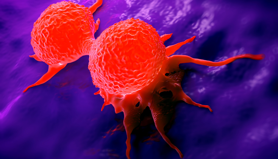

The majority of breast cancers begin in the lining of a breast milk duct and, if caught early, are highly curable. When these malignancies become invasive, they burst through a thin matrix surrounding the duct called the basement membrane and spread to adjacent tissue, making therapy more difficult. Stanford researchers revealed a novel physical mechanism that breast cancer cells exploit to break out and become invasive in a recent report published on Nov. 13 in Nature Materials. They discovered that, in addition to known chemical means of eroding the basement membrane, cancer cells cooperate together to physically distort and tear through the barrier.
“When this invasion process has been studied, the focus has typically been on single cells,” by courtesy of Ovijit Chaudhuri, an associate professor of mechanical engineering and bioengineering. “However, we do know that the invasion is collective in nature, with groups of cells cooperating to break through the basement membrane.” Our research has revealed how cells collaborate to break through the basement barrier, increasing our fundamental understanding of this important stage in cancer progression.”
A mechanical force model
Previous study has shown that individual cancer cells can release enzymes called proteases that tear down some of the basement membrane, but treatments that suppress proteases have not prevented cancer cells from escaping. Chaudhuri and his colleagues reasoned that there must be additional mechanisms at work, so they created a novel three-dimensional model to examine breast cancer and the basement membrane.
“What we’re really looking at are the mechanical forces involved, which is a different perspective,” explained Julie Chang, a PhD student in Chaudhuri’s lab and one of the paper’s lead authors. “The current paradigm is that cells use chemicals to degrade through the basement membrane, but we show that this physical aspect is just as important.”
The researchers created a 3D hydrogel that resembles the qualities of breast tissue as well as produced cellular structures called acini that resemble a breast duct and are surrounded by their own basement membrane. scientists labeled the basement membrane with fluorescent markers so that scientists could detect and measure any distortion as malignant cells interacted with it. And what they discovered astounded them.
The cancer cells trapped within the acini expanded together, forcing the basement membrane to stretch like a balloon. This stretching process stretched and weakened the basement membrane, allowing cancer cells near the membrane to exert additional forces to open holes and escape.
“The cells are expanding and increasing their volume in unison, and then locally pulling to tear up the basement membrane,” said Aashrith Saraswathibhatla, a postdoctoral researcher in Chaudhuri’s group and co-lead author on the article. “This collective volume expansion hasn’t been predicted before – no one was thinking about it.”
The researchers discovered that major findings from their 3D model were similar with what had been seen in individuals with invasive breast cancer. They also collaborated with colleagues at the University of Pennsylvania, who were able to corroborate their findings using computational modeling, indicating that the physical forces involved might hypothetically allow cells to burst through the basement membrane.
From novel mechanisms to novel therapies
Understanding how cancer cells interact with one another and the mechanical forces they exert could lead to novel therapeutic ways to prevent invasion. It could also help researchers estimate which patients with pre-invasive breast cancer are most likely to have their cancer break out and spread, which could lead to more focused treatments.
While this effort is only the first step toward these possibilities, the researchers are making progress in this area. They are looking into how cancerous cells physically interact with surrounding breast tissue once they break through the basement membrane, as well as delving deeper into understanding the basement membrane itself – looking into both mechanical and structural features – to gain a better understanding of why some tumors become invasive while others do not.
“This global volume expansion mechanism represents a new insight into how the breast tumors become invasive, borne out of the development of a 3D culture model that allowed us to visualize the invasion process,” says Chaudhuri. “It highlights the emergent behaviors arising in groups of cells acting together that enable cancer invasion.”
more recommended stories
 Nanoplastics in Brain Tissue and Neurological Risk
Key Takeaways for HCPs Nanoplastics are.
 AI Predicts Chronic GVHD Risk After Stem Cell Transplant
Key Takeaways A new AI-driven tool,.
 Red Meat Consumption Linked to Higher Diabetes Odds
Key Takeaways Higher intake of total,.
 Pediatric Crohn’s Disease Microbial Signature Identified
Key Points at a Glance NYU.
 Nanovaccine Design Boosts Immune Attack on HPV Tumors
Key Highlights Reconfiguring peptide orientation significantly.
 High-Fat Diets Cause Damage to Metabolic Health
Key Points Takeaways High-fat and ketogenic.
 Acute Ischemic Stroke: New Evidence for Neuroprotection
Key Highlights A Phase III clinical.
 Statins Rarely Cause Side Effects, Large Trials Show
Key Points at a Glance Large.
 Anxiety Reduction and Emotional Support on Social Media
Key Summary Anxiety commonly begins in.
 Liquid Biopsy Measures Epigenetic Instability in Cancer
Key Takeaways Johns Hopkins researchers developed.

Leave a Comment