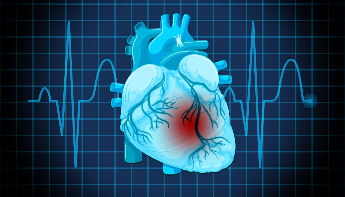
STUDY: Enhanced Imaging of Cardiac Micro-Vessels for Undiagnosed Chest Pain
Undiagnosed chest pain and heart problems could be better assessed with the use of a novel imaging approach that examines cardiac micro-vessels. This technology has been tested on patients.
To create sub-millimeter resolution images of cardiac micro-vessels, researchers from the Department of Bioengineering and the Faculty of Medicine at Imperial College London collaborated with scholars from UCL. Four human patients underwent testing using the novel non-invasive imaging technology.
Large blood veins on the surface of the heart can be seen with current imaging methods. However, by imaging tiny micro-vessels within the heart muscle, this new technology may enable researchers to examine the physiology of the heart in more detail.
Physicians may find it easier to comprehend the role these vessels play in cardiovascular disorders such as microvascular coronary disease and cardiomyopathies, as well as in unexplained chest pain, thanks to research published in Nature Biomedical Engineering.
Visualizing cardiac vessels is crucial for managing cardiovascular diseases, but there is a lack of understanding of how the blood flows within the small vessels of the heart. Our study images these vessels non-invasively in the highest resolution yet which, following further research, could help clinicians to manage these diseases.”- Professor Mengxing Tang, from the Department of Bioengineering at Imperial College London, and the corresponding author of the research
Seeing tiny vessels
For the heart to circulate blood throughout the body and provide tissues with oxygen while expelling waste and carbon dioxide, it must have efficient blood flow. On the other hand, aberrant blood flow from damaged cardiac vessels may result in tissue damage and heart failure.Four patients with hypertrophic cardiomyopathy (HCM) were used by the scientists to test the imaging method, which captured high-resolution images of the patients’ hearts’ cardiac micro-vessels and flow dynamics using ultrasonography and microbubbles.
Imaging the micro-vessels was difficult because of their size and the heart’s rapid motions, particularly at resolutions of less than a millimeter.
The method used by the researcher may be useful in assessing various heart problems. For instance, the method might be used by medical professionals to see anatomical abnormalities in individuals with cardiomyopathies and microvascular coronary disease, which would facilitate diagnosis and treatment and lead to better patient outcomes.
Professor Tang said: “This is the first time we demonstrated it is possible to image these vessels in such resolution, which has never been done before in humans. This has opened up a wide range of opportunities to study heart physiology and observe different diseases and conditions non-invasively and safely.”
Co-author and cardiologist Professor Roxy Senior, from the National Heart and Lung Institute at Imperial College London, said, “For the first time this technique allows direct visualization of the very small heart muscle vessels which when diseased give rise to chest pain which can be not only debilitating but may also lead to death. Because at present these vessels can be assessed only by indirect means the condition can be misdiagnosed.”
Upcoming uses and additional study
Despite the initial success, the research is still in its early phases, and additional studies with a larger number of patients would be necessary to fully comprehend the technique’s therapeutic and research significance. Image quality may also be improved by more research.
Professor Tang stated: “We are optimistic about what our new technique could bring to cardiovascular patient healthcare.”
In collaboration with oncologists, cardiologists, radiologists, breast surgeons, and other physicians, Professor Tang is also investigating the possible applications of super-resolution ultrasound technology for the assessment of a variety of other disorders.
He said: “This far-reaching research would never happen without collaboration between interdisciplinary teams of engineering and clinical sciences researchers.”
For more information: Transthoracic ultrasound localization microscopy of myocardial vasculature in patients, Nature Biomedical Engineering, doi.org/10.1038/s41551-024-01206-6
 Chronic Xanthan Gum Alters Gut Microbiota and Colon Health
Key Points Chronic xanthan gum consumption.
 Calcific Aortic Valve Stenosis Linked to Gum Disease Bacteria
Key Points Researchers identified a potential.
 Drug Delivery Nanoparticles Studied with New AF4-SANS Tool
Key Points Researchers developed the first.
 Gut-Friendly Diet Linked to Lower CHD Mortality Risk
Key Summary A higher Dietary Index.
 Parkinson’s Disease Risk Linked to Short Sleep, Study Finds
Key Points A large Chinese cohort.
 Cancer Risk And Allergic Diseases Linked in New Study
Key Points A large meta-analysis of.
 Lifestyle Behaviors and Mood Linked in Daily Life Study
Key Points A 70-day diary study.
 Cat Fleas Linked to Murine Typhus Risk in Texas Study
Key Points Researchers detected Rickettsia typhi.
 Pediatric Surgical Safety Reduces Serious Surgical Events
Key Points Pediatric operating room safety.
 England Obesity Prevalence Climbs to 30%, Lancet Study
Key Points A nationwide study of.
Leave a Comment