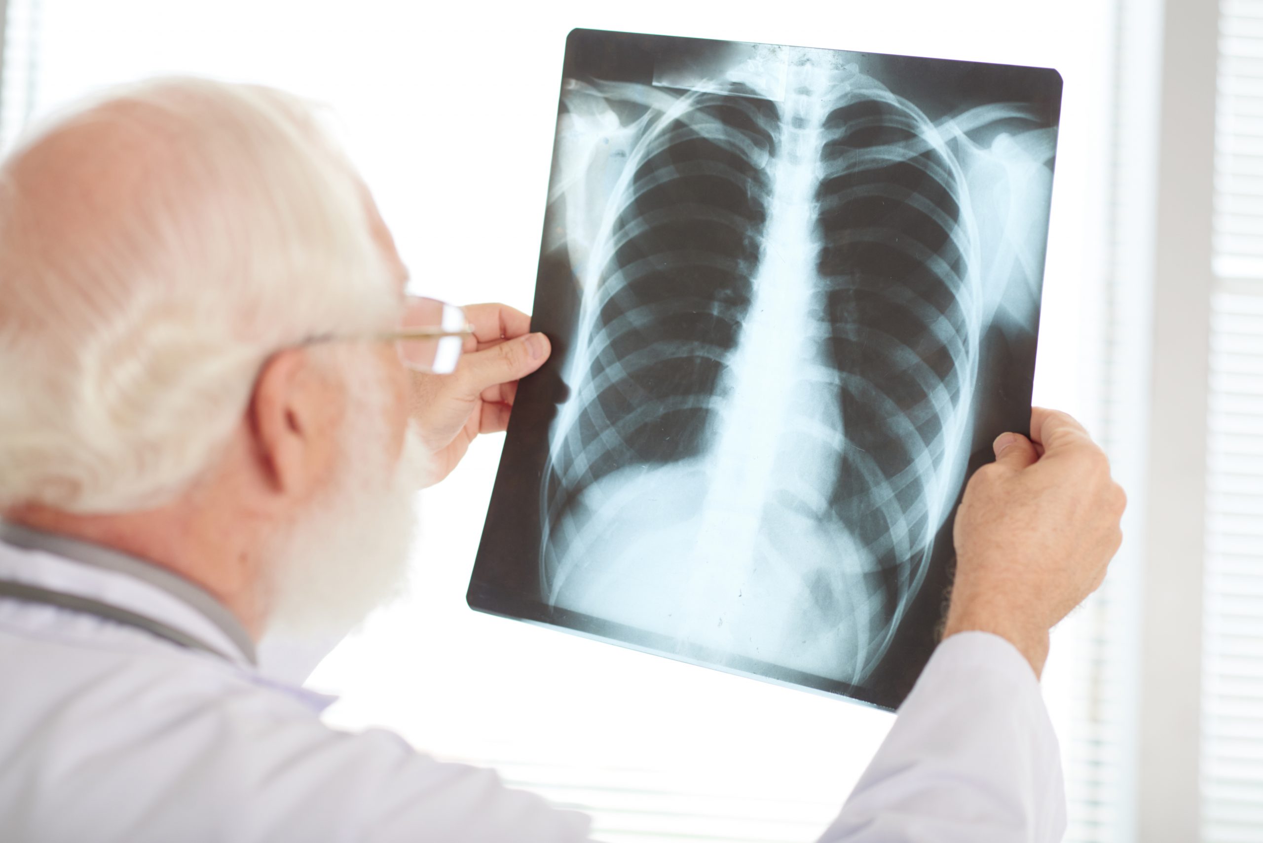

According to a recent study, an imaging technique that is currently only utilized in research labs could potentially be expanded for use in hospitals and clinics in order to detect early-stage lung disease.
Using a model of the human chest created by Duke University, scientists from the KTH Royal Institute of Technology in Stockholm examined the feasibility of utilizing a procedure called phase-contrast X-ray imaging on human lungs.
They claimed that the smallest airways—those measuring less than 2 mm—and any disease-related blockages could be seen using phase-contrast chest radiography. These are features that traditional radiography doesn’t capture, according to the study’s co-lead authors, KTH Royal Institute of Technology researchers Ilian Häggmark and Kian Shaker.
The Proceedings of the National Academy of Sciences published the findings that the researchers presented.
In research labs, soft tissue samples the size of a centimeter can only be imaged using the equipment now in use. But, according to Häggmark, the study unequivocally demonstrates that more can be done using phase-contrast X-ray imaging provided the technological requirements for clinical usage can be built.
According to Häggmark, chest radiography now used in hospitals and clinics serves a significant role in the early detection of respiratory diseases but is essentially constrained by the way it produces images.
When screening for disorders like asthma or chronic obstructive lung disease, he claims that the promising phase-contrast technique utilized in the study could reveal tiny pathological alterations that are otherwise undetectable with standard X-ray imaging (COPD).
“Phase-contrast X-ray imaging can extract more information at higher resolution using the same amount of radiation dose as in conventional radiography,” Häggmark says.
In traditional radiography, the X-ray beam travels through the body and is absorbed in various tissues to varying degrees along the way. After the beam has passed through the body and been filtered, it is measured by a detector on the opposite side. Attenuation is the procedure responsible for creating the contrast that gives X-ray images their usefulness.
Each X-ray beam can be used to extract more data using the phase-contrast method. This is due to the fact that variations in the waveforms of X-rays that pass through a sample can be measured. The phase of an X-ray wave can change in relation to a reference wave at any time when it interacts with atoms and other structures.
In the human chest, this enhanced image reveals the boundaries of the tiny airways and bronchial walls with stronger contrast and better clarity. This image is created using phase information from the sample.
Moving the detector further away from the patient, according to Häggmark, is one of the secrets to the technique.
Development of equipment for imaging larger samples will take time, he says. “You need an X-ray source with both high power and a small emission spot,” he says. “Basically, you need bright X-ray sources.”
He says that promising developments are being carried out, but it will take time for this to reach testing for human use,” he says.
more recommended stories
 Phage Therapy Study Reveals RNA-Based Infection Control
Key Takeaways (Quick Summary) Researchers uncovered.
 Safer Allogeneic Stem Cell Transplants with Treg Therapy
A new preclinical study from the.
 AI in Emergency Medicine and Clinician Decision Accuracy
Emergency teams rely on rapid, accurate.
 Innovative AI Boosts Epilepsy Seizure Prediction by 44%
Transforming Seizure Prediction in Epilepsy Seizure.
 Hypnosis Boosts NIV Tolerance in Respiratory Failure
A New Approach: Hypnosis Improves NIV.
 Bee-Sting Microneedle Patch for Painless Drug Delivery
Microneedle Patch: A Pain-Free Alternative for.
 AI Reshapes Anticoagulation in Atrial Fibrillation Care
Understanding the Challenge of Atrial Fibrillation.
 Hemoglobin as Brain Antioxidant in Neurodegenerative Disease
Uncovering the Brain’s Own Defense Against.
 Global Data Resource for Progressive MS Research (Multiple Sclerosis)
The International Progressive MS Alliance has.
 AI Diabetes Risk Detection: Early T2D Prediction
A new frontier in early diabetes.

Leave a Comment