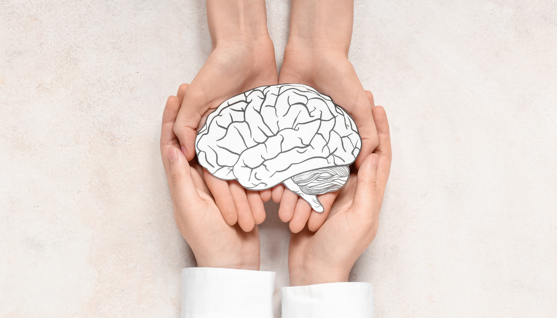
Functional Iron Deficiency in Parkinson’s Disease

Parkinson’s disease (PD) has long been associated with excess iron accumulation in the substantia nigra, reinforcing the belief that iron-driven oxidative stress and ferroptosis contribute to dopaminergic neuron loss. However, a recent perspective published in the Journal of Clinical Investigation challenges this dominant model, proposing that Parkinson’s disease may instead reflect iron starvation at the cellular level.
Researchers argue that while total brain iron may appear elevated on imaging, biologically usable iron is insufficient in vulnerable neurons. This concept, known as functional iron deficiency, suggests that iron is present but trapped in forms unavailable for critical cellular processes such as dopamine synthesis and mitochondrial metabolism.
Unexpected findings from clinical trials using deferiprone, a brain-penetrant iron chelator, have played a pivotal role in reshaping this narrative. Instead of improving outcomes, iron chelation worsened motor symptoms, particularly in untreated or early-stage Parkinson’s patients. These results raise an important clinical question: could iron removal further deprive neurons already struggling with iron-dependent functions?
Dopamine synthesis relies on tyrosine hydroxylase, an iron-dependent enzyme. Historical studies from the 1980s showed that iron supplementation improved motor symptoms and reduced reliance on dopaminergic therapy in some patients. Although these trials lacked modern controls, they align with current observations that iron availability, not excess, may limit neuronal function.
Advanced imaging techniques cannot differentiate between ferric (Fe³⁺) and ferrous (Fe²⁺) iron. Fe³⁺, often stored in ferritin or neuromelanin, is readily detected by MRI but is largely inert. In contrast, Fe²⁺ is essential for enzymatic activity and cellular energy production.
In Parkinson’s disease, iron may become sequestered due to lysosomal dysfunction, neuroinflammation, or glial trapping, creating the appearance of iron excess while neurons experience iron deprivation. Supporting evidence includes genetic NBIA disorders, manganism-related Parkinsonism, and animal models where impaired iron uptake leads to dopaminergic neuron loss.
This emerging framework challenges iron chelation as a universal strategy in Parkinson’s disease. Instead, targeted restoration of iron bioavailability, guided by disease stage and dopaminergic treatment status, may warrant further investigation. For clinicians and researchers, these insights highlight the need to reassess iron biology in neurodegeneration and its therapeutic implications.

 Tirzepatide Weight Loss: Fat Mass and Muscle Findings
Key points Tirzepatide was associated with.
 Coffee and Gut Microbiome: Brewing and Metabolic Effects
Key Points Coffee contains caffeine, chlorogenic.
 Low-Dose Radiation Therapy Shows Promise in Early Knee OA
Key Points Low-dose radiation therapy was.
 Oral Hormone Replacement Therapy Raises Blood Clot Risk
Key Points Oral hormone replacement therapy.
 GLP-1 Health Information: Quality, Completeness, & Engagement
Key Points A study assessed 128.
 DNA Damage Response May Drive Biological Aging
Key Points Chronic DNA damage response.
 Dietary Polyphenol Intake Associated With Lower MCI Odds
Key Points Older adults without mild.
 Maternal Diet Fails to Reduce Infant Egg and Peanut Allergy
Key Points A large Australian randomized.
 Psychedelic-Assisted Psychotherapy: Music in Treatment
Key Takeaways Psychedelic-assisted psychotherapy combines psychedelic.
 Cardiovascular Frailty Links Heart Health and Aging
Key Points Cardiovascular frailty may connect.
Leave a Comment