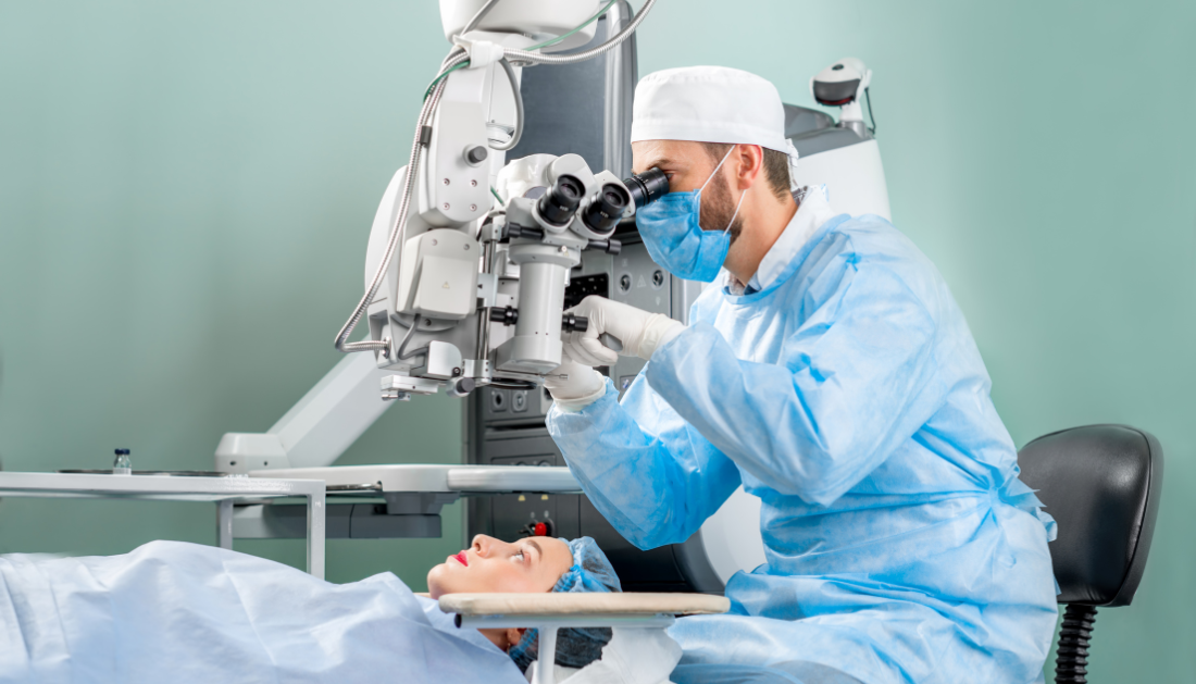

A surgical team at UNC Hospitals has successfully performed corneal neurotization, the only eyesight restoration surgical treatment for neurotrophic keratitis (NK), a rare eye condition that causes loss of sensation to the cornea and, if untreated, can lead to permanent vision loss. “It’s a life-changing procedure,” said Matthew Miller, MD, director of the UNC Facial Nerve Center and assistant professor of otolaryngology/head and neck surgery at the UNC School of Medicine. “It not only restores sensation in their eyes, but it can prevent and even reverse vision loss.”
Because the UNC team is the only one doing the surgery in North Carolina, South Carolina, and Virginia, this technique fills a critical access gap. In the United States, only 20 to 30 groups do corneal neurotization.
What exactly is Neurotrophic Keratitis?
The cornea is important for the general health of the eyeball. The cornea, like the windshield system of a car, protects the inner tissues of the eye and keeps the eye’s surface clean and moist. The cornea activates the blink reflex when debris drops on the surface or when it begins to dry out.
Blinking moisturizes the eye and removes unwanted dirt and bacteria. The cornea is also an important component of the eye’s optical system, and the purity and regularity of its surface are essential for good visual function.
Neurotrophic keratitis develops when the nerves that innervate the cornea are destroyed. It affects around 5,000 to 10,000 people in the United States. Trauma to the eye, viral infections, brain tumors, and brain surgery are the most prevalent causes of NK. When the cornea’s nerve supply is cut off, it is unable to keep itself moist or maintain a healthy architecture, resulting in ulcers, scarring, and, finally, permanent vision loss.
“If you lose your blink reflex, your cornea can become unhealthy, dry, and very inflamed,” stated Daniel Rubinstein, MD, ophthalmology assistant professor and oculofacial plastic surgeon on the case. “However, you can’t feel the pain or the dryness.” The outermost layer of the cornea can peel off over time, causing ulcers and scarring that compromises vision.”
The damaged eye must be removed in the most severe cases to prevent extensive infection and subsequent harm to other structures.
What Is the Process of Corneal Neurotization?
Simply defined, the surgery’s purpose is to restore sensation to the damaged cornea by transferring a neighboring healthy neuron.
Miller removes a sural nerve segment from the lower leg during the surgery. This will act as a graft or bridge to link a healthy nerve to a damaged cornea. Surgeons like to harvest this nerve because its sacrifice causes only a little region of numbness on the top of the foot.
Rubinstein then makes a small incision in each upper eyelid to gain access to the healthy supraorbital nerve, which is located immediately beneath the brow. The sural nerve transplant is then tunneled through incisions across the top face to link the afflicted eye to the healthy supraorbital nerve.
Miller painstakingly peels out the individual nerve fibers that make up the sural nerve using a sophisticated microscope. The fibers are then sewn to the injured cornea by Hussam Banna, MD, assistant professor of ophthalmology and cornea specialist. Miller completes the circuit under the microscope by stitching the other end of the sural nerve to a healthy “donor” nerve.
After about six months, the patient may notice improved sensation and, eventually, vision. The mending process is slow, but it changes patients’ lives.
Miller believes that the surgery’s minimally invasive approach is very essential for patients since it lowers scarring on the face, numbness, and other potential adverse effects.
“It is important because these patients, especially patients with facial paralysis and neurotrophic keratitis, have gone through very traumatic events,” Miller said. “Our faces are our everyday lives.” The last thing you want to do is leave a visible scar to remind them of their painful experiences in the past.”
A Unique Procedure
The difficult technique necessitates close cooperation among operating room nurses, technical staff, coordinators, and, in this case, three surgeons.
Miller, Rubinstein, and Banna, all experts in their own right, joined together at UNC Hospitals’ Hillsborough site, which held all of the specific medical devices required for the surgery.
“It’s pretty rare to have three surgeons operating on the same case, coordinating, and simultaneously taking care of the same patient,” Rubinstein said. “This type of collaborative patient care is rather unique to UNC.” We are really grateful to the hospital system and the institution for providing such generous support in order for us to undertake such a unique, life-changing operation for these patients. We look forward to continue our work in order to provide this treatment to any patient who may benefit from it.”
more recommended stories
 Nanoplastics in Brain Tissue and Neurological Risk
Key Takeaways for HCPs Nanoplastics are.
 AI Predicts Chronic GVHD Risk After Stem Cell Transplant
Key Takeaways A new AI-driven tool,.
 Red Meat Consumption Linked to Higher Diabetes Odds
Key Takeaways Higher intake of total,.
 Pediatric Crohn’s Disease Microbial Signature Identified
Key Points at a Glance NYU.
 Nanovaccine Design Boosts Immune Attack on HPV Tumors
Key Highlights Reconfiguring peptide orientation significantly.
 High-Fat Diets Cause Damage to Metabolic Health
Key Points Takeaways High-fat and ketogenic.
 Acute Ischemic Stroke: New Evidence for Neuroprotection
Key Highlights A Phase III clinical.
 Statins Rarely Cause Side Effects, Large Trials Show
Key Points at a Glance Large.
 Anxiety Reduction and Emotional Support on Social Media
Key Summary Anxiety commonly begins in.
 Liquid Biopsy Measures Epigenetic Instability in Cancer
Key Takeaways Johns Hopkins researchers developed.

Leave a Comment