Groundbreaking research suggests retinal thickness as an early marker for Parkinson's neurodegeneration.
The degree of the disease was found to be significantly correlated with a notable thinning of the retinal layer in the study, which followed changes in Parkinson’s patients from 2015 to 2021. According to these results, eye exams may develop into a non-invasive method for estimating the intensity of Parkinson’s disease symptoms in the future, enabling more specialized approaches to therapy.
Important Details:
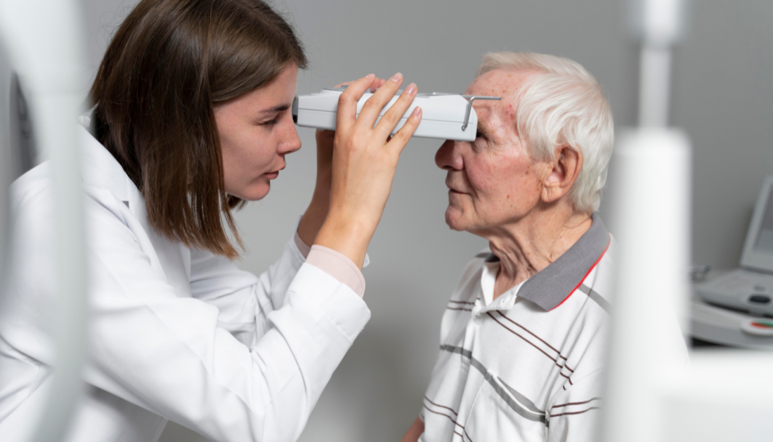
Retinal thickness was measured using optical coherence tomography in Parkinson’s patients, demonstrating that neurodegeneration in the retina occurs before cognitive and motor symptoms.
According to the study, an early-stage thinner retinal layer may be a sign of a more severe disease development and a quicker rate of cognitive impairment.
Regular eye exams may provide a non-invasive way to track and forecast Parkinson’s disease progression as a result of this research.
The Basque Country University is cited.
A method routinely used to perform ophthalmological tests can also be used to monitor the neurodegeneration that occurs in Parkinson’s patients, according to a study by the UPV/EHU and Biobizkaia. However, some aspects of this method still need confirmation for use in the clinical setting, and its resolution needs to be slightly improved.
During the investigation, it was discovered that retinal neurodegeneration most likely occurs before cognitive decline.
Patients who receive a diagnosis of Parkinson’s or similar neurological illness frequently inquire, “And now what? What will happen? What can be expected from the disease?”
“For neurologists, however, it is not possible to answer these questions precisely, as “the evolution of patients tends to be very varied: some experience no change over the years, while others end up with dementia or in a wheelchair,” outlined by researcher Ane Murueta-Goyena of the Neurosciences department at UPV/EHU.
Finding Parkinson’s patients who are at risk of cognitive impairment is becoming a significant difficulty, yet it is essential to advance clinical studies and develop more potent treatments.
Dr. Ane Murueta-Goyena sought to determine “whether the visual system can enable this deterioration to be predicted, in other words, what future the patient can expect within a few years.” in conjunction with the study team at Biobizkaia. For this, the retina’s thickness was utilized.
The retina is a membrane that is found at the rear of the eyeball. It is made up of multiple layers and is connected to the neurological system. Using optical coherence tomography, the thickness of the innermost layer of the retinas of a cohort of Parkinson’s patients was assessed during the investigation. This innovative approach not only sheds light on retinal health but also underscores the potential of ophthalmological techniques in understanding neurological conditions.
For more discussions on cutting-edge advancements in neurology, you can explore the Global Neurology Summit 2024.
Because this kind of tomography makes measurements that are precise, reproducible, and high-resolution, it is frequently utilized in ophthalmological examinations. Thus, between 2015 and 2021, the development of this retinal layer was examined and contrasted in individuals with and without Parkinson’s disease.
In a hospital in the UK, the findings of the examination of the retinal layer photographs of Parkinson’s patients were also validated.
The findings demonstrated that Parkinson’s sufferers had a notably thinner retinal layer. Additionally, it was noted that “during the initial phases of the disease it is in the retina where the greatest neurodegeneration is detected, and, from a given moment onwards, when the layer is already very thin, a kind of stabilizing of the neurodegeneration process takes place.”
“Retinal thinning and cognitive impairment do not occur simultaneously. The initial changes in the retina are more evident and then, over the years, patients are observed to worsen clinically in both cognitive and motor terms,” explained Murueta-Goya.
“In other words, the slower retinal layer thickness loss is associated with faster cognitive decline; this slowness is linked to greater severity of the disease.”
The findings are very important to the researcher. “We have obtained information on the progression of the disease, and the tool we are proposing is non-invasive and available at all hospitals.”
The outcomes must be verified globally, and “by slightly improving the resolution of the technology, we will be closer to validating the method for monitoring the neurodegeneration that takes place in Parkinson’s disease.” The researcher also disclosed that financing is essential and that they are carrying out the study on a different group of patients.
For more information: Association of retinal neurodegeneration with the progression of cognitive decline in Parkinson’s disease, npj Parkison’s disease, https://doi.org/10.1038/s41531-024-00637-x
 Calcific Aortic Valve Stenosis Linked to Gum Disease Bacteria
Key Points Researchers identified a potential.
 Drug Delivery Nanoparticles Studied with New AF4-SANS Tool
Key Points Researchers developed the first.
 Gut-Friendly Diet Linked to Lower CHD Mortality Risk
Key Summary A higher Dietary Index.
 Early Menopause Risk Higher in Rural Women: New Study
Key Points Early menopause affects more.
 Parkinson’s Disease Risk Linked to Short Sleep, Study Finds
Key Points A large Chinese cohort.
 Cancer Risk And Allergic Diseases Linked in New Study
Key Points A large meta-analysis of.
 Lifestyle Behaviors and Mood Linked in Daily Life Study
Key Points A 70-day diary study.
 Cat Fleas Linked to Murine Typhus Risk in Texas Study
Key Points Researchers detected Rickettsia typhi.
 Pediatric Surgical Safety Reduces Serious Surgical Events
Key Points Pediatric operating room safety.
 Blood Test May Improve Alzheimer’s Disease Prognosis
Key Points A new review suggests.
Leave a Comment