
Groundbreaking MRI scans reveal microstructural hypothalamic changes in young women, uncovering a potential biological foundation for eating disorders such as anorexia and obesity.
Hypothalamic Changes in Eating Disorders
The hypothalamus is a critical brain structure responsible for regulating hunger, mood, and hormonal balance. Despite its central role in feeding behaviors, it has been underexplored in female-focused neuropsychiatric research—particularly regarding why women are disproportionately affected by eating disorders during adolescence.
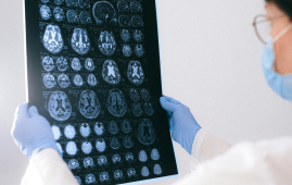
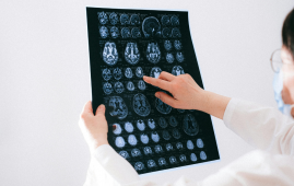
Traditional imaging techniques often fail to capture detailed changes in the hypothalamus. However, a new ultrahigh-resolution T1 MRI has allowed scientists to map this region with remarkable precision.
New Insights from High-Resolution MRI Imaging
A study published in the American Journal of Clinical Nutrition examined 44 young adult females—including those with anorexia nervosa, obesity, and normal weight—using 7T MRI to assess volumetric and cellular integrity of 50 hypothalamic regions.
Key findings included:
-
Significant structural differences in para- and periventricular nuclei among participants with eating disorders.
-
Volume increases in certain hypothalamic regions were linked to inflammation, potentially driving disordered eating patterns.
-
Hormonal fluctuations, especially in leptin and ghrelin levels, correlated with the severity of eating disorders.
Why Young Women Are at Greater Risk
The research indicates that larger hypothalamic subregions—likely due to inflammation—could disturb the brain’s regulatory control over food intake. This helps explain the increased vulnerability of young women to disorders like anorexia and obesity.
Moreover, the study found associations between specific hypothalamic regions and markers of mood, anxiety, and BMI, painting a complex neurobiological picture of eating disorders in females.
Toward Targeted Treatments Rooted in Hypothalamic Changes
This pioneering research opens doors to more personalized treatment strategies. Notably, GLP-1 receptor agonists, which affect the arcuate nucleus, may help manage disordered eating behaviors.
Future studies should explore whether hypothalamic alterations precede the onset of symptoms. Longitudinal and connectivity-based research could further clarify the role of the extended limbic and cortical networks involved.
Conclusion
With the aid of advanced MRI techniques, researchers are now able to visualize the hidden neurobiological factors contributing to eating disorders. These insights could transform early diagnosis, risk assessment, and targeted interventions—especially for young women most at risk.
For more information: Witte, A. V., & Sacher, J. (2025) Unraveling neural underpinnings of eating disorders in the female brain: Insights from high-field magnetic resonance imaging. The American Journal of Clinical Nutrition. 121(5), pp. 943-944. doi:10.1016/j.ajcnut.2025.02.027
more recommended stories
 Digital Heart Twins Show Promise for AF Ablation Precision
Key Summary Researchers developed patient-specific digital.
 Alzheimer’s Disease Study Questions Glucosamine Safety
Key Summary New research published in.
 WHO: Andes Hantavirus Not a COVID-19-Level Threat
Key Summary Andes hantavirus has drawn.
 Genetic Screening Project Launches for Brazilian Parents
Key Summary Brazil is launching the.
 CAR T-Cell Therapy Offers Hope for Highly Sensitized Patients
Key Summary Researchers at the University.
 Gut Keystone Bacteria: How Diet Shapes Microbiome Health
Key Takeaways Researchers are identifying “gut.
 Chronic Liver Disease in Europe Raises Public Health Alarm
Key Summary A new report in.
 CDC Nutrition Biomarkers Study Offers Public Health Insights
Key Points The upcoming CDC Nutrition.
 Macrophages Attack Live Melanoma Cells in Real Time
Key Takeaways Researchers at the Garvan.
 Facial Micromovements Linked to Accurate Pain Detection
Key Takeaways Researchers at Rutgers University–New.
Leave a Comment