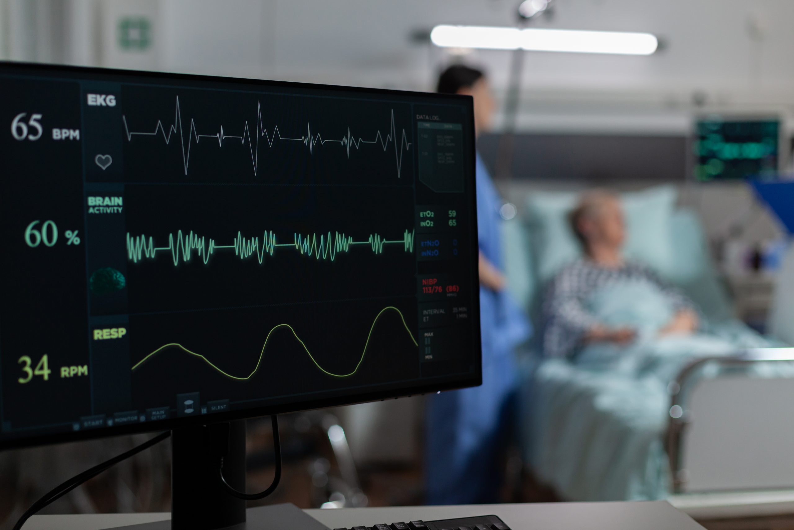
Image by DCStudio on Freepik

An automated screening technique for left ventricular (LV) systolic dysfunction is provided by a novel deep-learning application. The disorder significantly lowers the heart’s pumping ability and is connected with frequent hospitalizations and a double chance of mortality. With early detection and therapeutic administration, LV dysfunction can be avoided. However, detecting the disease before symptoms appear has proven impossible.
To solve this issue, Rohan Khera, MD, MS, and his colleagues at the Cardiovascular Data Science Lab (CarDS) Lab created a new artificial intelligence (AI)-based electrocardiogram (ECG) interpretation that is suitable for use worldwide. The findings were published in the journal Circulation on July 25.
Without an echocardiography or an MRI scan, a cardiologist cannot detect individuals with LV dysfunction. Cardiac imaging is required to diagnose LV systolic dysfunction, which is a weakening in the heart’s main chamber. Technology and available knowledge hinder broad screening for the disease. However, ECG is the most widely used cardiovascular diagnostic test in clinical practice worldwide.
The scientists used roughly 400,000 ECGs in their design, along with data on cardiac failure from imaging testing. The method was evaluated in a variety of formats using data from multiple clinics and hospitals in the United States, as well as a large community cohort in Brazil.
“We demonstrate that a simple photo or scanned image of a 12-lead ECG, the most well-recognized and easily obtained cardiac test, can provide key insights on cardiac structure and function disorders,” Khera said. “This opens up the possibility to finally bring a screening tool for such disorders that affect up to one in 20 adults globally.
“Their diagnosis is frequently delayed as advanced testing is either unavailable or only reserved for those with symptomatic disease. Now we can identify these patients with a simple web-based or smartphone application,” said Khera. A version of such an application accompanies the paper and is hosted by the CarDS Lab for demonstration.
“Our approach creates a super-reader of ECG images—identifying signatures of LV systolic dysfunction, which the human eye cannot accurately decipher,” said Veer Sangha, the first author of the study, a member of the CarDS Lab, and a Rhodes Scholar.
“Our AI tool allows early diagnosis and treatment and also identifies those at future risk of developing LV dysfunction,” said Khera. “The findings represent our ongoing effort to make application of AI-driven advanced ECG inference accessible.”

 Coffee and Gut Microbiome: Brewing and Metabolic Effects
Key Points Coffee contains caffeine, chlorogenic.
 Low-Dose Radiation Therapy Shows Promise in Early Knee OA
Key Points Low-dose radiation therapy was.
 Oral Hormone Replacement Therapy Raises Blood Clot Risk
Key Points Oral hormone replacement therapy.
 GLP-1 Health Information: Quality, Completeness, & Engagement
Key Points A study assessed 128.
 DNA Damage Response May Drive Biological Aging
Key Points Chronic DNA damage response.
 Maternal Diet Fails to Reduce Infant Egg and Peanut Allergy
Key Points A large Australian randomized.
 Psychedelic-Assisted Psychotherapy: Music in Treatment
Key Takeaways Psychedelic-assisted psychotherapy combines psychedelic.
 Cardiovascular Frailty Links Heart Health and Aging
Key Points Cardiovascular frailty may connect.
 Laser Blended Vision: Simple Test Predicts Success
Key Points A simple phoropter-based test.
 Pink Noise Boosts Brain Waste Clearance During Sleep
Key Points Pink noise delivered during.
Leave a Comment