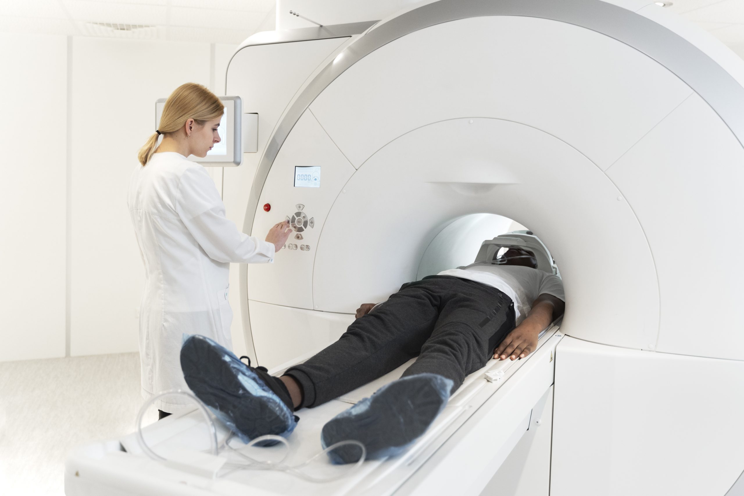

The majority of the approximately 250,000 Americans diagnosed with acute pulmonary embolism (PE) in emergency rooms each year are hospitalized. However, according to a new Michigan Medicine study published in JAMA Network Open, certain patients with PE, a blood clot in one or more pulmonary arteries, may be hospitalized needlessly due to computed tomography (CT Scan) imaging results rather than clinical risk factors.
According to the Pulmonary Embolism Severity Index, or PESI score, around 40% of the patients in the research had low-risk pulmonary embolism. Roughly half of the low-risk individuals had CT imaging abnormalities that doctors deemed “concerning,” and these patients did just as well in the hospital as those whose CT scan revealed no such issues.
“The results of this study are going to come as a surprise to most emergency physicians, who have been taught for years to regard large, centrally located, or ‘saddle,’ PEs as true medical emergencies,” said senior author Colin Greineder, M.D., Ph.D., an assistant professor of emergency medicine and pharmacology at University of Michigan Medical School.
“In fact, our findings suggest that CT findings alone might not confer as much risk as we thought. In our population, if the patient’s pulse, rate of breathing, oxygen level and other clinical factors indicated low risk, then ‘concerning’ CT findings by themselves were not associated with worse clinical outcomes.”
Between late 2016 and the end of 2019, researchers examined data from over 800 patients at U-M Health who were diagnosed with acute pulmonary embolism in the adult emergency department.
Patients were separated into low- and high-risk groups based on the PESI score, with the low-risk group further subdivided based on the existence of worrying CT scan findings. Large and centrally positioned PEs, blood clots on both sides of the lung, indications of heart strain due to the blockage, and accompanying infarction or death of a portion of lung tissue were among these.
Within a month of arriving at the ER, 18% of patients with high risk PESI scores died, but none of the low-risk patients with alarming CT imaging findings died. These individuals also seldom required intensive care, and their hospital stays were no longer than those of low-risk patients with no “concerning” CT imaging findings.
“Clinical outcomes were similar, but we found plenty of evidence that emergency physicians are managing these patients differently,” Greineder said. The rate of discharge and home management was about four times lower in low risk patients with concerning CT imaging findings, and they were more likely to have bedside ultrasounds performed in the emergency department and echocardiograms while in the hospital.
The study also looked into how frequently emergency physicians at U-M Health used the PE Response Team, a multidisciplinary group designed to make quick, shared decisions on the management of patients with intermediate and high risk PE.
Activation of the response team was nearly four times more common in low-risk patients with concerned CT imaging findings, despite their low PESI scores.
A strong evidence base has been established to support safe outpatient management of pulmonary embolism over the last two decades, but adoption by emergency medicine clinicians has been slow and continues to be limited, according to co-author Geoffrey Barnes, M.D., M.Sc., associate professor of cardiology-internal medicine at U-M Medical School.
“We have tools like the Pulmonary Embolism Severity Index and biomarkers that allow for appropriate management of these patients, but they do not typically include those ‘concerning’ CT findings. Our report suggests that these imaging findings may be an important barrier to safe discharge.”
According to the researchers, more investigation and validation are needed to ensure that these findings are repeated in different hospital systems and patient demographics.
“Like other groups, we are already working to overcome other barriers to outpatient management of low-risk PE,” said first author Connor O’Hare, M.D., an emergency medicine resident at U-M Health. “If our current findings are reproduced in other studies and hospital systems, then we will need effective implementation strategies to overcome the perceived risk associated with ‘concerning’ CT imaging findings in otherwise low-risk patients.”
more recommended stories
 Red Blood Cells Improve Glucose Tolerance Under Hypoxia
Key Takeaways for Clinicians Chronic hypoxia.
 Nanoplastics in Brain Tissue and Neurological Risk
Key Takeaways for HCPs Nanoplastics are.
 AI Predicts Chronic GVHD Risk After Stem Cell Transplant
Key Takeaways A new AI-driven tool,.
 Red Meat Consumption Linked to Higher Diabetes Odds
Key Takeaways Higher intake of total,.
 Pediatric Crohn’s Disease Microbial Signature Identified
Key Points at a Glance NYU.
 Nanovaccine Design Boosts Immune Attack on HPV Tumors
Key Highlights Reconfiguring peptide orientation significantly.
 High-Fat Diets Cause Damage to Metabolic Health
Key Points Takeaways High-fat and ketogenic.
 Acute Ischemic Stroke: New Evidence for Neuroprotection
Key Highlights A Phase III clinical.
 Statins Rarely Cause Side Effects, Large Trials Show
Key Points at a Glance Large.
 Anxiety Reduction and Emotional Support on Social Media
Key Summary Anxiety commonly begins in.

Leave a Comment