image by peoplecreations on Freepik
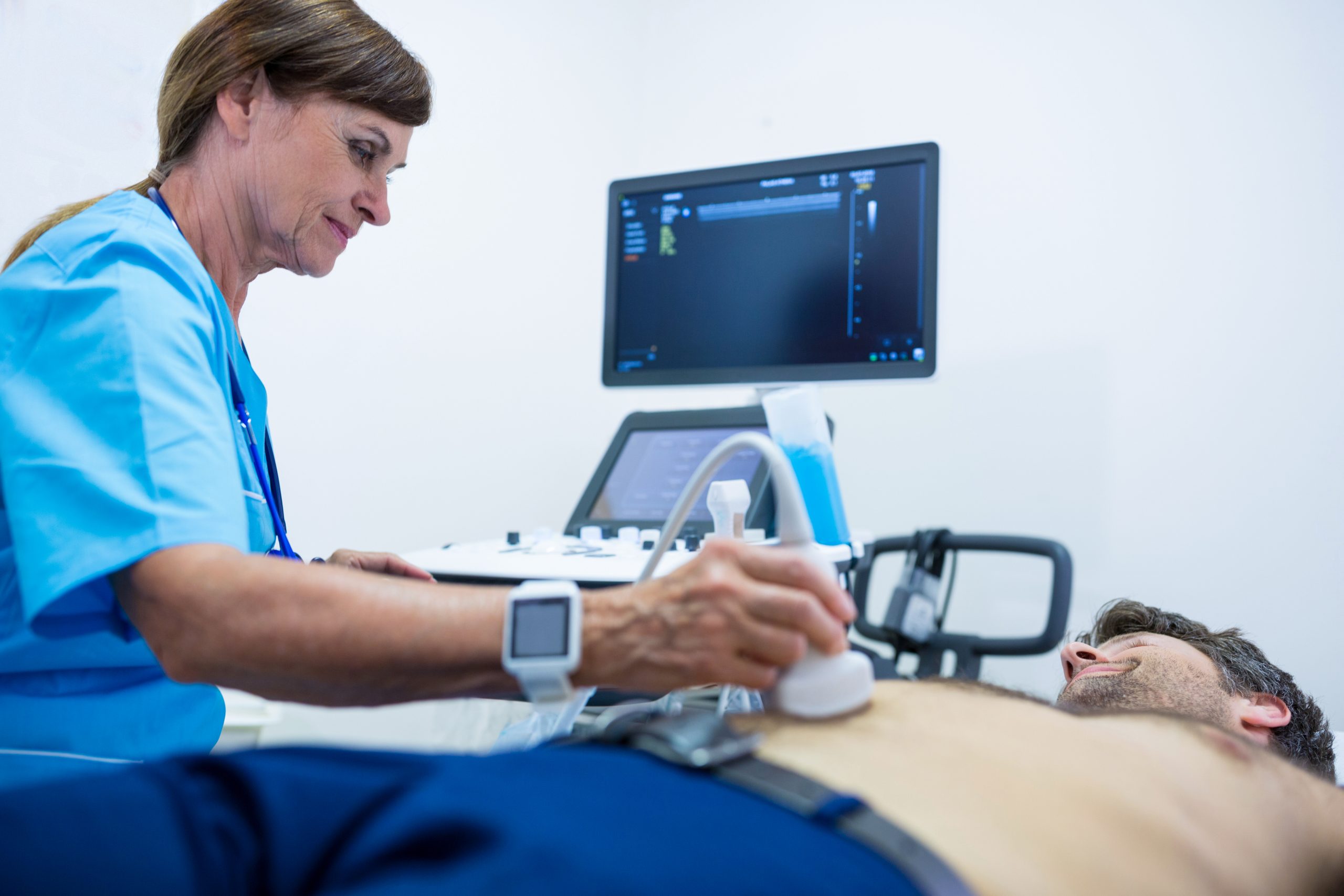
The majority of graft failures after heart transplantation are caused by anomalies such as significant coronary artery disease. As donors with more stringent requirements for heart transplantation, such as advanced age and pre-existing heart problems, become available, rigorous screening for congenital defects has become critical. Invasive coronary angiography is a critical screening tool for detecting coronary artery disease (CAD), a disorder marked by cholesterol deposits in the arteries of the heart. However, practical issues restrict its utility, therefore it is utilized by less than one-third of donors who are at risk of developing CAD. A new heart preservation approach known as “ex situ heart perfusion” (ESHP) has been developed to address this constraint. ESHP allows clinicians to monitor the performance of the heart and screen it for any problems outside the body by permitting the flow of oxygenated nutrients to the heart via blood arteries. However, coronary angiography during ESHP has been linked to heart injury and primary graft failure. Alternative coronary imaging technologies are thus necessary to detect aberrant blood flow in donors’ hearts and establish their transplantability.
According to the Journal of Biomedical Optics (JBO), a team of researchers led by Professor Elise Colin of Paris Saclay University in France has developed a non-invasive optical approach for evaluating coronary blood circulation in donor’s hearts during ESHP. The technology makes use of a recently discovered speckle imaging technique called Laser Speckle Orthogonal Contrast Imaging (LSOCI), which was created to detect the numerous scattering of moving red blood cells. The researchers increased LSOCI’s imaging capability to observe tiny blood veins in the heart in this investigation. Thus, the suggested approach evaluates blood flow in the heart by employing a polarimetric filtering procedure that suppresses surface scattering. As a result, the time-varying speckle patterns used in imaging are mostly produced by repeated scattering on moving red blood cells inside peripheral arteries.
“The optical technique allows high-resolution imaging of the entire peripheral vasculature of the heart in real-time,” says Colin.
To put this strategy to the test, the researchers created a clinical model of coronary circulation on donor’s hearts prior to transplantation. They then generated and analyzed rapidly varying speckle patterns with a laser and a camera set on an adjustable arm fixed above the perfusion module, which contained the donor’s heart.
To solve the difficulties in monitoring vasculature caused by heartbeat, the researchers improved the methodology with a method known as Multi-Period-Enhanced Signal-to-Noise Ratio (MPE-SNR). They collected a series of photos throughout time to create a set of frames depicting the vasculature at various heart locations. The other photos were then used to optimize each image in this sequence to reduce noise and enhance details.
The newly developed imaging technique enables precise visualization of blood circulation. In the future, this technique can potentially identify myocardial perfusion abnormalities that indicate underlying heart conditions, such as coronary artery disease.
According to Amy Oldenburg, Professor at the University of North Carolina at Chapel Hill and JBO Associate Editor, “The non-contact technique, based on laser speckle, provides imaging of blood circulation within donor’s hearts and has important implications for identifying hearts that are safe and suitable for transplantation.”
 Phage Therapy Study Reveals RNA-Based Infection Control
Key Takeaways (Quick Summary) Researchers uncovered.
 Safer Allogeneic Stem Cell Transplants with Treg Therapy
A new preclinical study from the.
 AI in Emergency Medicine and Clinician Decision Accuracy
Emergency teams rely on rapid, accurate.
 Innovative AI Boosts Epilepsy Seizure Prediction by 44%
Transforming Seizure Prediction in Epilepsy Seizure.
 Hypnosis Boosts NIV Tolerance in Respiratory Failure
A New Approach: Hypnosis Improves NIV.
 Bee-Sting Microneedle Patch for Painless Drug Delivery
Microneedle Patch: A Pain-Free Alternative for.
 AI Reshapes Anticoagulation in Atrial Fibrillation Care
Understanding the Challenge of Atrial Fibrillation.
 Hemoglobin as Brain Antioxidant in Neurodegenerative Disease
Uncovering the Brain’s Own Defense Against.
 Global Data Resource for Progressive MS Research (Multiple Sclerosis)
The International Progressive MS Alliance has.
 AI Diabetes Risk Detection: Early T2D Prediction
A new frontier in early diabetes.
Leave a Comment