Image by Freepik
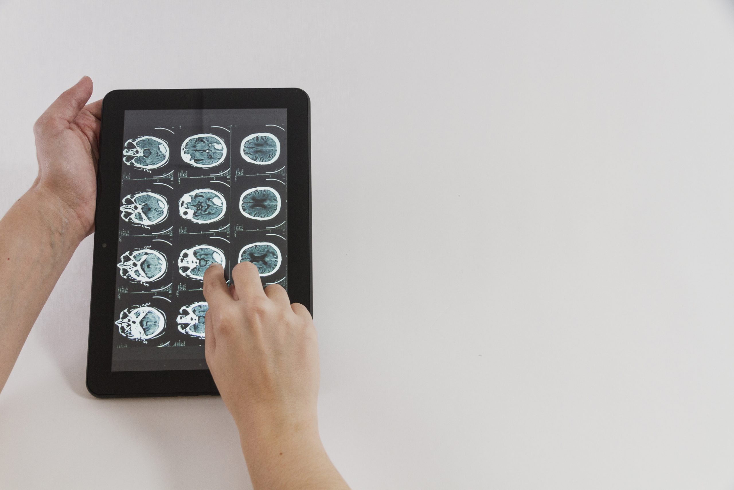
The University Hospital Bonn (UKB) neonatology team used mobile magnetic resonance imaging to carry out the first-ever study of kids getting ECMO therapy. (MRI). Extracorporeal membrane oxygenation (ECMO), as the process is also known, entails oxygenating the blood outside of the body. The results of the successful, ground-breaking research using the mobile MRI on the first four pediatric ECMO patients have been released in Critical Care.
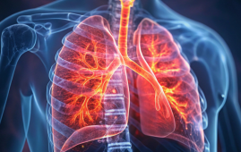
Critically ill patients are those who require ECMO treatment in addition to a conservative ventilator. Lung collapse, heart failure, or infection are possible causes. Only at specialized treatment facilities, like the Children’s ECMO Center of the UKB, where they are carefully monitored, can children who need this special procedure be treated. Both infants and older kids are treated here with ECMO therapy.
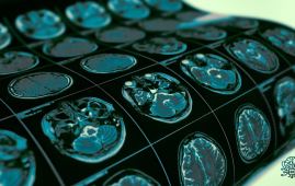
“It is often necessary for this sensitive group of patients, even during ECMO therapy, to have an MRI of the brain to check the relevant structures in the brain. However, transport to a fixed device is unfortunately not possible,” says Prof. Sabir. Last August, a grant from the Bill Gates Foundation enabled him to purchase a mobile MRI, which is being used at the UKB for the first time in Germany to clinically test diagnostics on premature and newborn infants. So far, it has only been used for research purposes in London.
The mobile MRI is a ground-breaking new advancement in the optimization of diagnostics for newborn patients and has already been in use at the UKB for more than half a year.
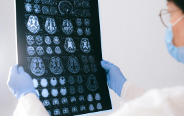
Since then, 25 kids have undergone mobile MRI scans at UKB, with the youngest weighing only 450 grams and the eldest being 10 years old. The mobile MRI was used for regular exams as well as for additional abnormality diagnosis, such as after asphyxia. (oxygen deficiency at birth). For each of the children who underwent the examination, a comparison image was obtained in the permanently installed normal-field MRI at the UKB in order to assess the image quality of the mobile low-field MRI.
“We were more than satisfied with the results. Although the image quality of the mobile MRI is not as high-resolution as that of a fixed device, the image data are ideal for emergency diagnostics and, above all, can be retrieved immediately. Among other things, we were able to detect brain hemorrhages, strokes or acute changes, such as the accumulation of cerebrospinal fluid, in the children examined so far and initiate the appropriate therapies immediately,” says Prof. Sabir.
ECMO patients, however, pose a special challenge to the treatment team in connection with MRI diagnostics. “While adults have a tube inserted in the groin area to transport blood during ECMO therapy, children often have access at the neck. The patients have to be moved very carefully and the tube at the neck may only be moved minimally,” explains Prof. Sabir.
However, only MRI imaging allows for accurate diagnoses, such as those for brain hemorrhages. As a result, the neonatology and pediatric critical care team was able to show that mobile MRI imaging can be successfully completed in four pediatric ECMO patients.
The patients included an infant, a child aged two, a child aged nine, and a child aged ten. Using the mobile MRI, one of the kids had a significant brain hemorrhage that was identified and managed right away.
“The new findings prove that the scan can be performed safely. We obtained meaningful MRI images of the brain without changing the position of the neck cannula and without compromising the children’s safety status. This represents an immense success for future MRI examinations of newborns and larger children who can only survive through the use of ECMO therapy,” said Prof. Andreas Müller, Director of the Department of Neonatology at UKB.
 Phage Therapy Study Reveals RNA-Based Infection Control
Key Takeaways (Quick Summary) Researchers uncovered.
 Safer Allogeneic Stem Cell Transplants with Treg Therapy
A new preclinical study from the.
 AI in Emergency Medicine and Clinician Decision Accuracy
Emergency teams rely on rapid, accurate.
 Innovative AI Boosts Epilepsy Seizure Prediction by 44%
Transforming Seizure Prediction in Epilepsy Seizure.
 Hypnosis Boosts NIV Tolerance in Respiratory Failure
A New Approach: Hypnosis Improves NIV.
 Bee-Sting Microneedle Patch for Painless Drug Delivery
Microneedle Patch: A Pain-Free Alternative for.
 AI Reshapes Anticoagulation in Atrial Fibrillation Care
Understanding the Challenge of Atrial Fibrillation.
 Hemoglobin as Brain Antioxidant in Neurodegenerative Disease
Uncovering the Brain’s Own Defense Against.
 Global Data Resource for Progressive MS Research (Multiple Sclerosis)
The International Progressive MS Alliance has.
 AI Diabetes Risk Detection: Early T2D Prediction
A new frontier in early diabetes.
Leave a Comment