Intrinsic Cardiac Nervous System Controls Heart Function in Mice

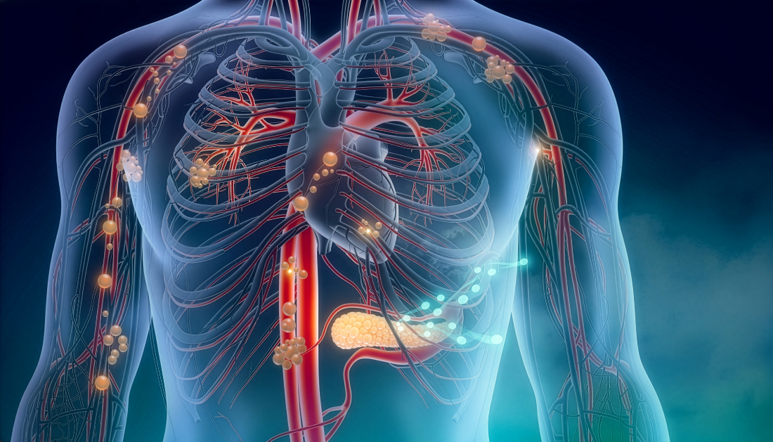
A new study published in Cell has revealed that the intrinsic cardiac nervous system (ICNS) contains two specialized nerve cell populations that perform distinct yet complementary roles in maintaining cardiac stability in mice. The findings provide new insight into how the heart’s internal neural network regulates routine cardiovascular function while protecting against electrical instability during extreme stress.
For cardiologists, electrophysiologists, cardiovascular researchers, and other healthcare professionals, the research offers a clearer understanding of the cellular organization of the ICNS and highlights potential directions for precision neuromodulatory therapies. While the discoveries are limited to mouse models, they establish an important framework for future translational research into human cardiac disorders.
Using single-cell RNA sequencing, genetic engineering, advanced imaging, and cell-specific neuromodulation, researchers mapped thousands of intrinsic cardiac neurons and identified four molecular clusters. Two dominant neuron populations, Npy-positive and Ddah1-positive neurons, demonstrated distinct anatomical connections and physiological functions.
The Npy-positive neurons primarily received parasympathetic (vagal) input and extensively innervated the sinoatrial (SA) node, atrioventricular (AV) node, ventricular tissue, and the aortic root. These neurons regulated heart rate and supported coronary perfusion by influencing aortic-root mechanics. When researchers selectively removed these neurons, mice developed fatal cardiac failure, highlighting their essential role in maintaining normal cardiac performance.
Meanwhile, Ddah1-positive neurons were concentrated within posterior cardiac ganglia and primarily received sympathetic input. Their nerve fibers projected to the pulmonary veins, pulmonary artery, and left atrial regions, which are closely associated with atrial arrhythmias. These neurons played a critical role in preserving electrical stability during severe physiological and psychological stress.
To evaluate the protective role of these neurons, investigators exposed mice to physical restraint, prolonged heat stress, and pharmacologically induced sympathetic overactivation using epinephrine and caffeine.
Animals lacking Ddah1-positive neurons became significantly more susceptible to malignant arrhythmias and sudden cardiac arrest under stress. In contrast, selective activation of these neurons improved survival during sympathetic overstimulation, suggesting they function as an intrinsic safeguard against electrical collapse.
The study also demonstrated that each neuronal population receives distinct autonomic inputs and innervates distinct cardiac regions, indicating that the ICNS comprises highly specialized neural circuits rather than a uniform network.
Current cardiac neuromodulation approaches, including radiofrequency ablation, electrical stimulation, and cryoablation, generally affect broad areas of the cardiac nervous system without distinguishing between neuron subtypes. These findings suggest that future therapies could focus on specific ICN populations to improve therapeutic precision while minimizing unintended effects.
Although the investigators identified similarities between these cardiac neurons and certain gastrointestinal neuron populations, additional research is needed to define their precise mechanisms, downstream signaling pathways, and functional relevance in humans.
The authors conclude that future studies should determine whether the molecular organization identified in mice is conserved in the human heart. If confirmed, these specialized neuronal circuits may provide promising targets for precision therapies designed to preserve cardiac rhythm, improve autonomic regulation, and reduce the risk of stress-induced arrhythmias.

 Gamma Oscillations Restored in Alzheimer’s Disease Mice
Key takeaways Researchers restored parvalbumin (PV).
 Body Roundness Index Linked to Higher Heart Attack Risk
Key findings at a glance Study.
 Visual Impairment Linked to Higher Dementia Risk
Key findings at a glance A.
 Epilepsy Drugs May Interact With Hormonal Contraceptives
Key points: Nearly one-third of people.
 Night Shift Work Alters Key Circadian Gene Expression
A recent study published in Frontiers.
 Dietary Polyphenol Intake Associated With Lower MCI Odds
Key Points Older adults without mild.
 Cardiovascular Frailty Links Heart Health and Aging
Key Points Cardiovascular frailty may connect.
 Pink Noise Boosts Brain Waste Clearance During Sleep
Key Points Pink noise delivered during.
 Maternal Illness Linked to Epigenetic Fetal Brain Changes
Maternal illness during pregnancy may influence.
 Rett Syndrome: Carotid Body Linked to Breathing Dysfunction
Key Points Rett syndrome is associated.
Leave a Comment