
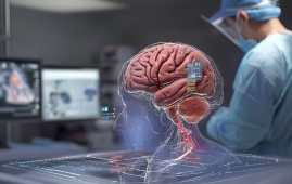
Image by kjpargeter on freepik
Researchers have found a way to screen for genetic alterations in dangerous brain tumors in under 90 seconds using artificial intelligence. This might potentially speed up the detection and treatment of gliomas, according to a study.
An AI-based diagnostic screening system called DeepGlioma was created by a group of neurosurgeons and engineers at Michigan Medicine in collaboration with researchers from New York University, the University of California, San Francisco, and other institutions. This system uses rapid imaging to analyze tumor specimens obtained during an operation and find genetic mutations more quickly.
The recently created approach successfully identified mutations used by the World Health Organization to establish molecular subgroups of the condition in a study of more than 150 individuals with diffuse glioma, the most prevalent and lethal primary brain tumor. The average accuracy was over 90%.
The results are published in Nature Medicine.
“This AI-based tool has the potential to improve the access and speed of diagnosis and care of patients with deadly brain tumors,” said lead author and creator of DeepGlioma Todd Hollon, M.D., a neurosurgeon at the University of Michigan Health and assistant professor of neurosurgery at U-M Medical School.
Given that the advantages and hazards of surgery vary among patients with brain tumors depending on their genetic make-up, molecular categorization is becoming more and more important in the diagnosis and treatment of gliomas. In fact, patients with a particular kind of diffuse glioma called astrocytomas can live an additional five years on average after having their entire tumor removed.
However, diffuse glioma molecular testing availability is constrained and not consistently offered at facilities that treat patients with brain malignancies. When it is accessible, according to Hollon, it may take days or even weeks to get findings. “Barriers to molecular diagnosis can result in suboptimal care for patients with brain tumors, complicating surgical decision-making and selection of chemoradiation regimens,” Hollon said.
Before DeepGlioma, there was no way for doctors to tell diffuse gliomas apart during surgery. The technology, which was developed in 2019, combines deep neural networks with the stimulated Raman histology optical imaging technique, which was also created at the University of Michigan, to examine brain tumor tissue in real time.
“DeepGlioma creates an avenue for accurate and more timely identification that would give providers a better chance to define treatments and predict patient prognosis,” Hollon said.
Even with optimal standard-of-care treatment, patients with diffuse glioma face limited treatment options. The median survival time for patients with malignant diffuse gliomas is only 18 months.
While the development of medications to treat the tumors is essential, fewer than 10% of patients with glioma are enrolled in clinical trials, which often limit participation by molecular subgroups. Researchers hope that DeepGlioma can be a catalyst for early trial enrollment.
Progress in the treatment of the most deadly brain tumors has been limited in the past decades- in part because it has been hard to identify the patients who would benefit most from targeted therapies,” said senior author Daniel Orringer, M.D., an associate professor of neurosurgery and pathology at NYU Grossman School of Medicine, who developed stimulated Raman histology. “Rapid methods for molecular classification hold great promise for rethinking clinical trial design and bringing new therapies to patients.”
 Phage Therapy Study Reveals RNA-Based Infection Control
Key Takeaways (Quick Summary) Researchers uncovered.
 Safer Allogeneic Stem Cell Transplants with Treg Therapy
A new preclinical study from the.
 AI in Emergency Medicine and Clinician Decision Accuracy
Emergency teams rely on rapid, accurate.
 Innovative AI Boosts Epilepsy Seizure Prediction by 44%
Transforming Seizure Prediction in Epilepsy Seizure.
 Hypnosis Boosts NIV Tolerance in Respiratory Failure
A New Approach: Hypnosis Improves NIV.
 Bee-Sting Microneedle Patch for Painless Drug Delivery
Microneedle Patch: A Pain-Free Alternative for.
 AI Reshapes Anticoagulation in Atrial Fibrillation Care
Understanding the Challenge of Atrial Fibrillation.
 Hemoglobin as Brain Antioxidant in Neurodegenerative Disease
Uncovering the Brain’s Own Defense Against.
 Global Data Resource for Progressive MS Research (Multiple Sclerosis)
The International Progressive MS Alliance has.
 AI Diabetes Risk Detection: Early T2D Prediction
A new frontier in early diabetes.
Leave a Comment