Image by brgfx on Freepik
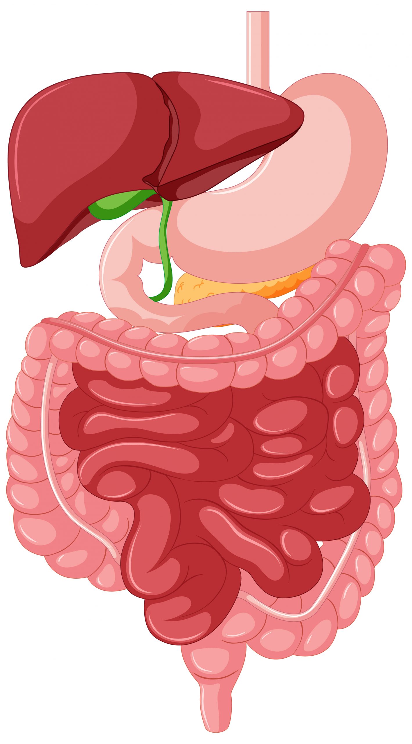
Food can be detected and located in the esophagus by sensory cells in the vagus nerve. Their signals aid in the delivery of food to the stomach. According to a team led by Carmen Birchmeier at the Max Delbrück Center, signal failure causes swallowing difficulties. They published their findings in the journal Neuron.
Swallowing disorders can have a variety of reasons, and they are more common in older persons. However, neurological illnesses such as multiple sclerosis and Parkinson’s disease, as well as some drugs, can cause food to move abnormally from the mouth to the stomach. Malnutrition, weight loss, and dehydration are all possible outcomes.
A team led by Professor Carmen Birchmeier, who directs the Developmental Biology/Signal Transduction Lab at the Max Delbrück Center in Berlin, has now studied the swallowing process in greater depth.
The researchers describe how sensory cells of the vagus nerve respond to mechanical inputs in the esophagus and cause involuntary muscle movement, a phenomenon known as esophageal peristalsis, in the journal Neuron. The vagus nerve, one of the 12 cranial nerves, sends information to the brain regarding the health of internal organs. The findings of the team’s investigation could eventually lead to better therapies for swallowing difficulties.
Swallowing on Camera
“Modern methods of single-cell sequencing made our work possible,” explains Birchmeier. “Using the sequencing data, we constructed genetic models that allowed us to study the functions of the sensory neurons in the vagal ganglia in more detail.” Ganglia are a group or “node” of neuronal bodies in the peripheral nervous system.
The researchers began by labeling neurons to determine which organs they innervate. The researchers then investigated if and how they responded to mechanical stimuli in the esophagus. Finally, they turned off the cells to see how this affected swallowing. Dr. Teresa Lever of the University of Missouri School of Medicine in Columbia, Missouri, created a technique that enables researchers to use video fluoroscopy to record swallowing in freely functioning, non-anesthetized mice in real time.
Beyond a Tube
“When mice lost the neurons that provide information about mechanical stimuli in the esophagus, they lost the ability to reflexively perform the appropriate muscle movements that transport food to the stomach, and they quickly lost weight,” says lead author Dr. Elijah Lowenstein, who earned his Ph.D. working on this study in Birchmeier’s team. He’s now a researcher at Harvard Medical School in Boston. The weight loss, says Lowenstein, shows that the neurons play a key role in bodily homeostasis.
“So, the esophagus isn’t just a tube that connects the mouth to the stomach,” he says. “It uses mechanosensory feedback to fulfill its function.” Birchmeier adds that without these cells in the vagus nerve, food gets stuck in our esophagus. In some of the mice, it actually flowed back into the throat.
A Comprehensive Molecular Atlas for All
“Our work can now help develop better treatments for swallowing disorders. One option would be to pharmacologically activate the mechanoreceptors we identified,” says Birchmeier. She also wants to use the genetic models to determine the functions of other vagal sensory neurons—such as those that control the lungs or aorta.
These neurons probably play a crucial but as-yet unknown role in the development of certain respiratory diseases, or cardiovascular diseases such as hypertension,” she says. Other researchers can also participate in these projects, as Birchmeier and her team have developed a molecular atlas for all vagal neurons in mice. The atlas is freely available online.
 Drinking Water With Meals Linked to Higher Food Intake
Key Points Summary A new Appetite.
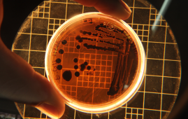
 Cyclosporiasis Risk: Who Faces the Highest Threat?
Key Summary Cyclosporiasis cases are increasing.
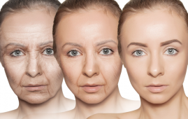 EP2 Receptor Research Opens New Path for Healthy Aging
Key Points Researchers identified the EP2.
 Obesity-Associated Leukemia Linked to Inflammation
Key Takeaways Researchers identified how obesity.
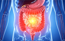 Chronic Xanthan Gum Alters Gut Microbiota and Colon Health
Key Points Chronic xanthan gum consumption.
 Calcific Aortic Valve Stenosis Linked to Gum Disease Bacteria
Key Points Researchers identified a potential.
 Drug Delivery Nanoparticles Studied with New AF4-SANS Tool
Key Points Researchers developed the first.
 Gut-Friendly Diet Linked to Lower CHD Mortality Risk
Key Summary A higher Dietary Index.
 Parkinson’s Disease Risk Linked to Short Sleep, Study Finds
Key Points A large Chinese cohort.
 Cancer Risk And Allergic Diseases Linked in New Study
Key Points A large meta-analysis of.
Leave a Comment