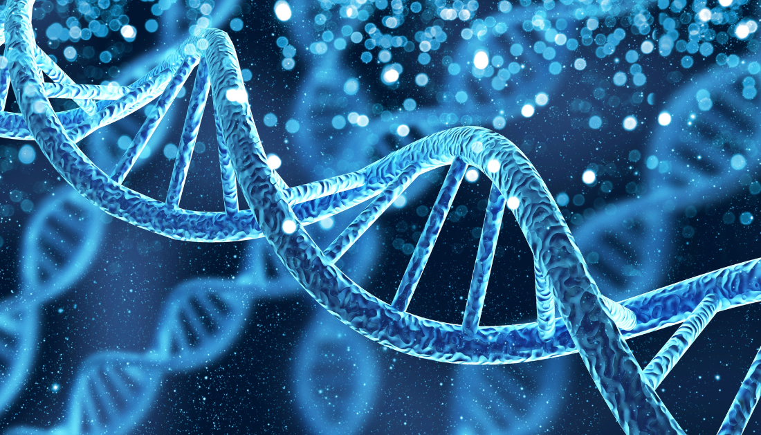

The same team reported in a recent study that a gene previously linked to autism spectrum disorder (ASD) by UT Southwestern Medical Center researchers appeared to play a critical role in directing cells in the brain’s hippocampus toward their ultimate identities. The discoveries, which were published in Science Advances, may potentially lead to new treatments for the common neurodevelopmental disease.
“This study is one of the few that provides a mechanistic understanding of autism spectrum disorder,” said senior author Maria Chahrour, Ph.D., Associate Professor of Neuroscience and Psychiatry and an Investigator in the Peter O’Donnell Jr. Brain Institute at UT Southwestern.
According to the Centers for Disease Control and Prevention, one out of every 36 children in the United States has ASD. Dr. Chahrour emphasized the importance of genetics in ASD by stating that twin studies indicate roughly 90% heritability. Although hundreds of genes related with ASD have been found, she added, it is unclear how these genes contribute to the illness.
Dr. Chahrour and her colleagues discovered an ASD-associated gene named KDM5A in 2020, demonstrating that individuals with mutations in this gene exhibit ASD, a lack of speech, intellectual incapacity, and other symptoms. Although KDM5A encodes a chromatin regulator (a protein that influences how DNA is packed in cells and whether other genes are expressed), the mechanism underlying its significance in ASD was unknown.
Dr. Chahrour and her colleagues investigated the variety of cell types in a mouse model in which this gene has been deleted, knowing that chromatin regulators control cell identity or how cells evolve into particular types. They focused on the hippocampus, the brain’s learning and memory area, whose structure and function are disrupted in autism spectrum disorder (ASD).
The hippocampus is made up of four major cell types: excitatory neurons, inhibitory neurons, glia, and endothelial cells, which are further subdivided into 24 subtypes. The researchers analyzed more than 105,000 nuclei using a technique known as single-nuclei RNA sequencing to compare the populations of cell types present in the hippocampus of mice with KDM5A versus animals lacking the gene, or “knockouts.”
Their findings revealed distinct differences in four excitatory neuron subtypes and two inhibitory neuron subtypes. Some of these cell types increased in number, others dropped, and one converted to a new subtype within its class in mice lacking KDM5A, indicating that KDM5A plays an important role in determining cell identity during development.
A closer inspection at the cells in the hippocampi of KDM5A knockout animals revealed that they were more developed, with abnormally more and longer branching cells than in KDM5A animals. Many of the damaged cells were found in the hippocampus’s CA1, which is important for maintaining social memories, or recollections of interactions with people. Changes in cell types can cause excitation and inhibition imbalances, and abnormalities in cellular development can harm hippocampal circuits and lead to hippocampus dysfunction, accounting for some of the symptoms associated with ASD, according to Dr. Chahrour.
Dr. Chahrour, an Associate Professor in the Eugene McDermott Center for Human Growth and Development and the Center for the Genetics of Host Defense at UTSW, said the findings advance her lab’s critical mission of understanding the molecular mechanisms underlying ASD and related neurodevelopmental conditions.
more recommended stories
 Acute Ischemic Stroke: New Evidence for Neuroprotection
Key Highlights A Phase III clinical.
 Statins Rarely Cause Side Effects, Large Trials Show
Key Points at a Glance Large.
 Anxiety Reduction and Emotional Support on Social Media
Key Summary Anxiety commonly begins in.
 Liquid Biopsy Measures Epigenetic Instability in Cancer
Key Takeaways Johns Hopkins researchers developed.
 Human Antibody Drug Response Prediction Gets an Upgrade
Key Takeaways A new humanized antibody.
 Pancreatic Cancer Research: Triple-Drug Therapy Success
Key Summary Spanish researchers report complete.
 Immune Cell Epigenome Links Genetics and Life Experience
Key Takeaway Summary Immune cell responses.
 Dietary Melatonin Linked to Depression Risk: New Study
Key Summary Cross-sectional analysis of 8,320.
 Chronic Pain Linked to CGIC Brain Circuit, Study Finds
Key Takeaways University of Colorado Boulder.
 New Insights Into Immune-Driven Heart Failure Progression
Key Highlights (Quick Summary) Progressive Heart.

Leave a Comment