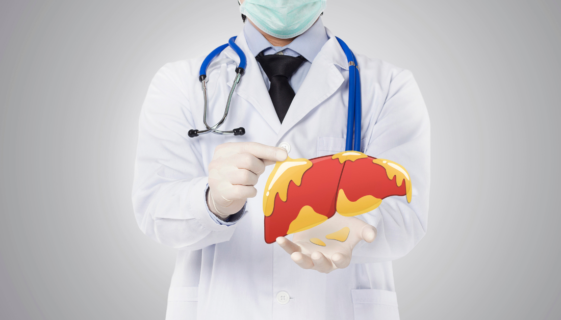
Study: Markers of insulin resistance associated with non-alcoholic fatty liver disease in non-diabetic population
Researchers investigate whether insulin- and non-insulin-based insulin resistance (IR) markers can predict the risk of non-alcoholic fatty liver disease (NAFLD) in obese and non-obese adults with no history of diabetes or hepatitis in a recent study published in the journal Scientific Reports.
Background
The current diagnostic test for non-obese NAFLD, liver biopsy, is invasive. Alternative methods like MRI and CT are costly. While ultrasound imaging is affordable, it lacks sensitivity to mild steatosis and varies in interpretation by physicians. There is a lack of suitable diagnostic methods for atypical non-obese NAFLD. Previous studies have produced conflicting results regarding the association between IR markers and NAFLD risk. However, IR markers may aid in the precise early detection of NAFLD in non-obese patients due to their link with liver fibrosis in NAFLD patients without diabetes.
About the Study
In this study, 2,148 subjects were recruited between 2021 and 2023. Information on their gender, age, current and past medical history, and medication history was collected. The participants were categorized as non-obese or obese based on their computed body mass index (BMI), with values of less than 25 kg/m2 and exceeding 25 kg/m2, respectively.
Venous blood samples were taken after the participants fasted for at least 10 hours. Four non-insulin-based insulin resistance (IR) markers were assessed: homeostatic model assessment of IR (HOMA-IR), triglyceride-glucose (TyG) index, TyG index with BMI (TyG-BMI), triglyceride/high-density lipoprotein cholesterol ratio (TG/HDL-c), and metabolic score for IR (METS-IR). The levels of these biomarkers were compared in predicting non-alcoholic fatty liver disease (NAFLD) in obese and non-obese individuals.
Logistic regression models were used to evaluate the relationship between IR markers and NAFLD risk. The IR marker values were divided into four quartiles.
The researchers assessed the predictive ability of IR markers for NAFLD using the area under the curve (AUC) and receiver operating characteristic (ROC) values. They also quantified the correlation between IR markers and NAFLD risk using the odds ratio (OR) and 95% confidence interval (CI), with a statistical significance threshold set at a p-value of less than 0.05.
Study Findings
Fasting plasma glucose (FPG), uric acid (UA), TG, aminotransferase (AST), alanine aminotransferase (ALT), and gamma-glutamyl transpeptidase (GGT) levels were considerably higher in the NAFLD group. Notably, the BMI of the NAFLD obese and non-obese categories was similarly significantly higher.
All five IR indicators, including HOMA-IR, TyG, TyG-BMI, TG/HDL-c, and METS-IR, had significantly greater odds ratios in the NAFLD group than in the non-NAFLD group. These levels frequently rose with rising quartile levels in the general study cohort, as well as in the obese and non-obese subgroups.
The AUC of TyG-BMI was highest in the non-obese category, implying that TyG-BMI had a stronger predictive value for NAFLD in non-diabetic and non-obese patients. The AUC of HOMA-IR was highest in the obese category, indicating that HOMA-IR is a stronger predictor of NAFLD in non-diabetic obese people. Furthermore, the AUC of each IR marker was greater than 0.5 with p-values less than 0.05, showing statistically significant and specific NAFLD predictive qualities.
Conclusion
The findings of the study back up previous studies of IR indicators being linked to an increased risk of NAFLD. Thus, utilizing TyG-BMI and HOMA-IR IR indicators to diagnose non-obese and obese NAFLD appears to be clinically meaningful, as they were associated with superior detection capacities than the other IR markers.
Although the researchers could not explain why this difference exists, they hypothesize that because TyG-BMI is derived based on body fat distribution utilizing FPG, TG, and BMI measurements, it has superior diagnostic value in non-obese individuals. Several studies, however, demonstrate that normal BMI in non-obese patients is still an independent risk factor for NAFLD.
For more information: Markers of insulin resistance associated with non-alcoholic fatty liver disease in non-diabetic population. Scientific Reports 13(1); 1-8. doi:10.1038/s41598-023-47269-4
 One Health Summit: WHO Leads Global Health Response
Key Highlights Global leaders convened in.
 Prenatal Smoking Raises Risk of Child Mental Disorders
Key Summary Prenatal smoking is associated.
 Physical Activity Guidelines Gap: Walking is Insufficient
Quick Summary Walking remains the most.
 Breast Cancer Risk Rises with Aging Tissue Changes
Key Highlights A 3-million-cell atlas reveals.
 Type 2 Diabetes Risk Rising in Genetically Susceptible
Quick Summary Rising type 2 diabetes.
 Female Microbiome Shaped by Diet, Stress, Unhealthy Lifestyle
Quick Summary Lifestyle factors significantly influence.
 Transcatheter Valve-in-Valve Improves Mitral Outcomes
Key Highlights Transcatheter mitral valve-in-valve (mVIV).
 Stroke Rehabilitation: Early High-Intensity Therapy Findings
Key Highlights High-intensity therapy within 2.
 TRPM8 Cold Sensation Mechanism Explained for Pain Care
Quick Summary TRPM8 ion channel converts.
 Gum Recession from Snus Confirmed, Caries Risk Debated
Key Highlights Snus use is strongly.
Leave a Comment