Image by macrovector on Freepik
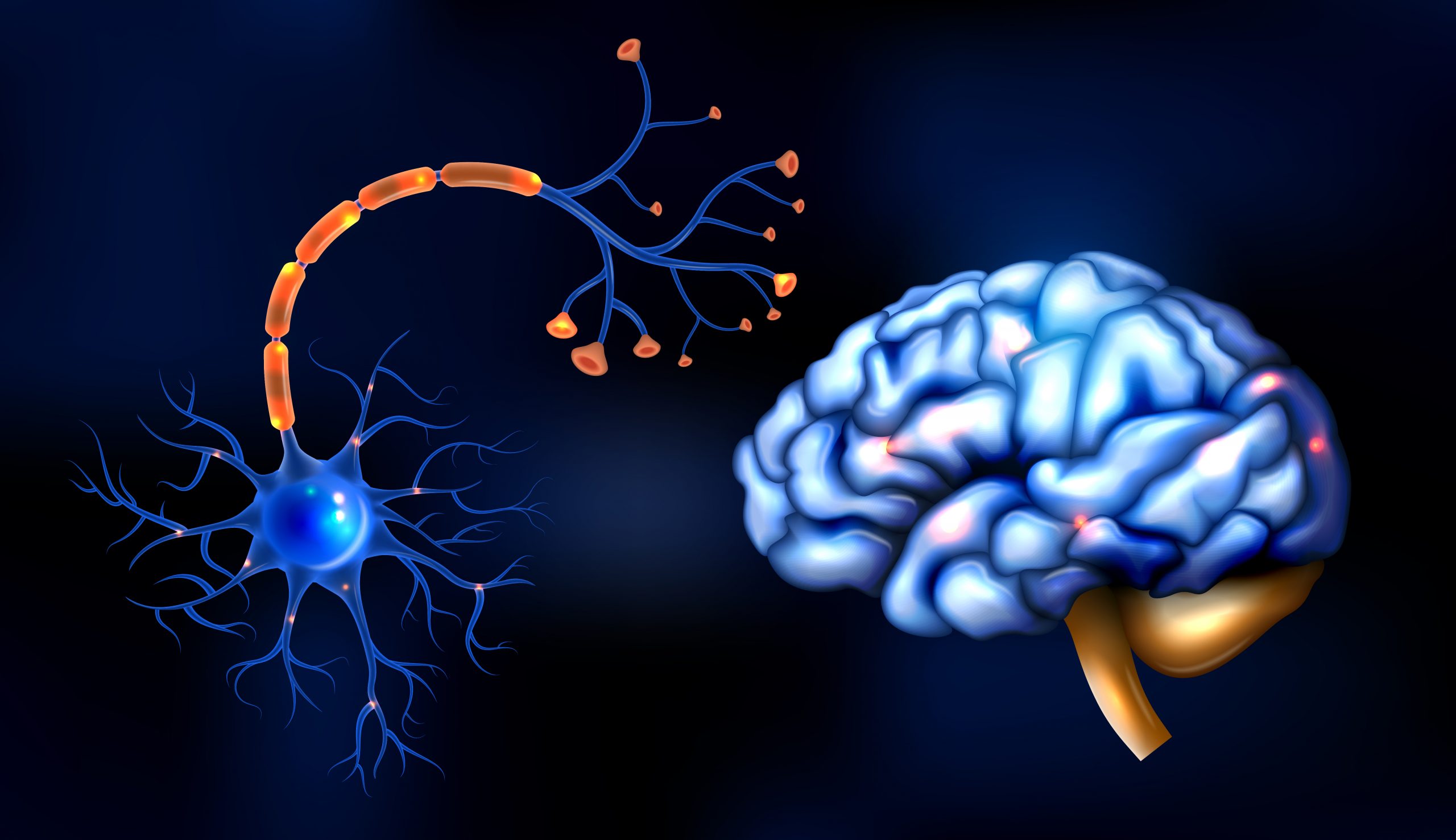
Scientists have discovered the chemical receptor structure that is essential for brain development and function.
Because of their importance in brain function, Type A GABA receptors are already targeted by pharmaceutical anesthetics, sedatives, and antidepressants. The study, which was published today in the journal Nature, exposes the dominant assemblies and states of the GABA receptor, which could lead to the development of new drugs that target a variety of medical problems more specifically.
“It is the main player that balances excitation and inhibition in the brain,” said lead author Chang Sun, Ph.D., a postdoctoral researcher in the Vollum Institute at Oregon Health & Science University. “It affects all aspects of brain function, from motor function, to memory and learning, and also emotion and anxiety.”
“Because the off switch is so crucial, GABA receptors are spread throughout the entire brain,” added senior author Eric Gouaux, Ph.D., senior scientist in OHSU’s Vollum Institute and an investigator with the Howard Hughes Medical Institute.
The receptor is defined by five-sided, or pentameric, assemblies derived from 19 distinct subunits, each of which gives rise to a vast number of clinically relevant configurations. In this example, researchers painstakingly separated native assemblies from mice before infusing them with commonly used medicines for sleeplessness and postpartum depression.
They were then able to see the receptor’s three major structural populations.
“This study shows the dominant assemblies and states of the GABA receptor,” Gouaux said. “That’s really the huge breakthrough — nobody had been able to figure out which of the hundreds of thousands of these assemblies are most highly populated.”
According to co-author Sarah Clark, Ph.D., a former postdoctoral fellow in the Gouaux lab and currently an assistant professor at Oregon State University, the discovery demonstrates the GABA receptor in its native state rather than tissue culture, as previously demonstrated. Researchers used cutting-edge cryogenic electron microscopy to disclose the structure in its natural state, as opposed to previous techniques that required crystallizing enormous amounts of similar molecules to generate a false representation of their original structure.
“We used a combination of cryo-EM as well as single-molecule microscopy technique, which allowed us to count the subunits in each pentameric complex,” she said.
Gouaux commended OHSU, as well as the Jennifer and Bernard Lacroute Endowed Chair in Neuroscience, for sponsoring this high-risk, high-reward research, as well as the Howard Hughes Medical Institute for providing persistent support over a three-year period that resulted in the discovery.
“This kind of work is difficult to fund because no one thinks it will work,” Gouaux said.
 Physical Activity Guidelines Gap: Walking is Insufficient
Quick Summary Walking remains the most.
 Breast Cancer Risk Rises with Aging Tissue Changes
Key Highlights A 3-million-cell atlas reveals.
 Type 2 Diabetes Risk Rising in Genetically Susceptible
Quick Summary Rising type 2 diabetes.
 Female Microbiome Shaped by Diet, Stress, Unhealthy Lifestyle
Quick Summary Lifestyle factors significantly influence.
 Transcatheter Valve-in-Valve Improves Mitral Outcomes
Key Highlights Transcatheter mitral valve-in-valve (mVIV).
 Stroke Rehabilitation: Early High-Intensity Therapy Findings
Key Highlights High-intensity therapy within 2.
 TRPM8 Cold Sensation Mechanism Explained for Pain Care
Quick Summary TRPM8 ion channel converts.
 Gum Recession from Snus Confirmed, Caries Risk Debated
Key Highlights Snus use is strongly.
 Hypertensive Disorders of Pregnancy: Role of Daily Activity
Key Points Summary Limiting sedentary time.
 Climate Change Drives Dengue Outbreaks Globally
Key Takeaways Extreme weather significantly increases.
Leave a Comment