Image on Freepik
Every cell in the body expends a tremendous amount of energy to maintain the proper ratio of water to essential electrolytes in order to live. Researchers at Oregon Health & Science University have created a method to map this action in the human brain and other organs in great detail using magnetic resonance imaging, or MRI, scanning. The development, known as metabolic activity diffusion imaging, or MADI, is creating new opportunities for the early detection of cancer and determining whether a tumor is reacting to therapy. Researchers will contrast MADI with positron emission tomography, or PET, which uses injected radioactive agents to generate images of cell energy production rates, in upcoming clinical trials involving patients with glioma brain tumors.
MADI is a new way to make images of metabolic activity within organs and tissues at high spatial resolution, and it’s totally noninvasive,” said inventor Charles Springer, Ph.D., professor with the OHSU Advanced Imaging Research Center. “In principle, this method could apply to almost any pathology. Right now, we are pushing it in the direction of cancer and neuroscience.”
In an animal model using rats, the OHSU researchers already have shown that MADI can detect and monitor brain tumors as effectively as PET, but without the need for injecting tracers or contrast agents of any kind.
“It tells us more about what’s going on inside cells in regard to ion transport, water transport, energy production, and so we think it will definitely be useful in cancer and other diseases,” said Martin Pike, Ph.D., associate professor with the OHSU Advanced Imaging Research Center, who is leading the glioma studies.
MADI also provides higher-resolution images than PET. “It can resolve regions of metabolic activity inside the tumor,” Springer said. “None of the current clinical methods used to map metabolic activity has the spatial resolution needed to measure variations in metabolism within any but the largest tumors.”
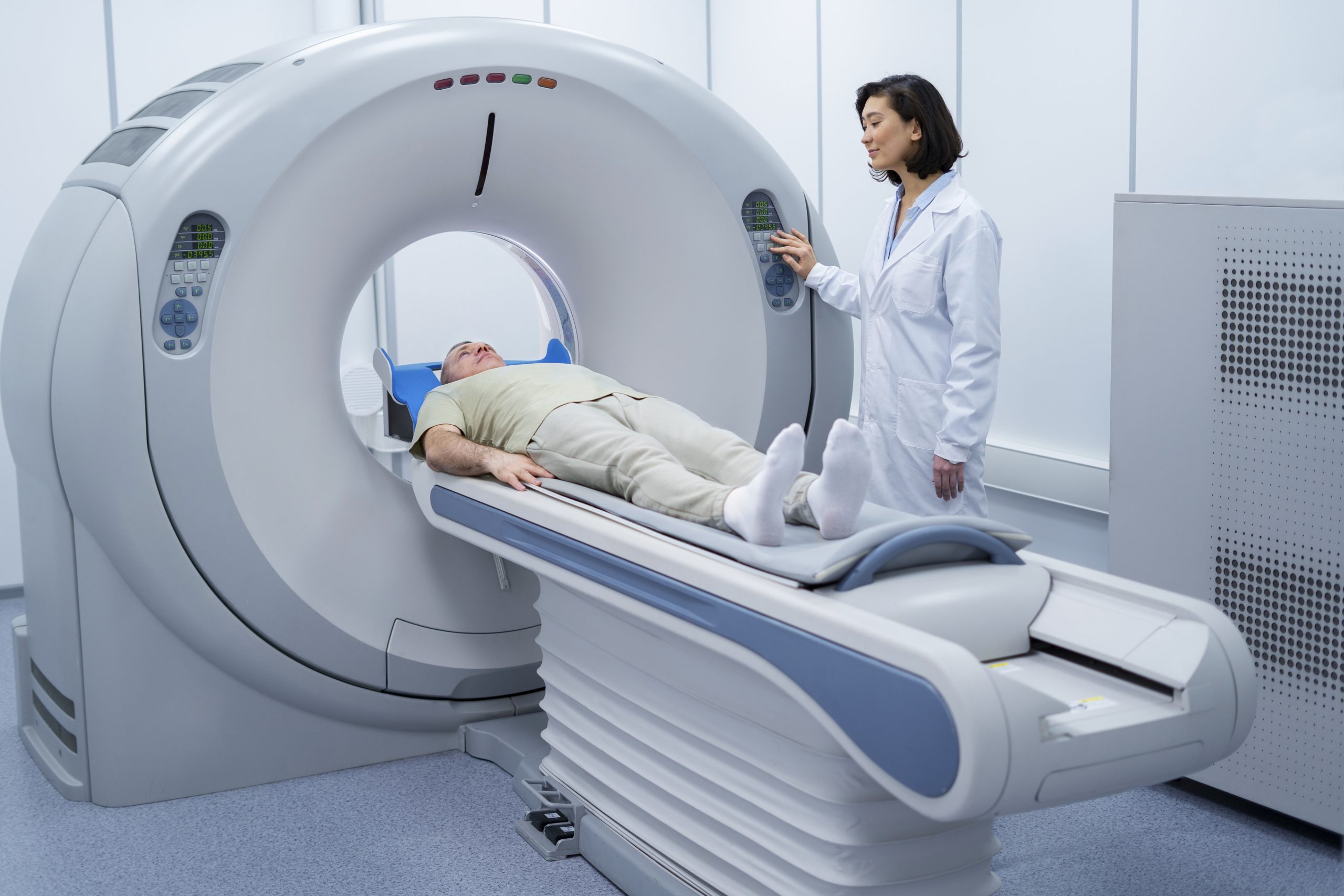
Associate professor of diagnostic radiology at the OHSU School of Medicine Ramon Barajas, M.D., who is working on the glioma research, points out that understanding how various tumor components function can be very helpful in making a diagnosis. A strong magnetic field is used by MRI to produce incredibly precise images of internal organs. The water droplets’ hydrogen atom nuclei align with the magnetic field as a result of the magnetic field. The radio wave pulses are then delivered by the MRI machine at a resonance frequency. Magnetized hydrogen nuclei respond by emitting radio waves once more, which causes signals to be detected by the MRI machine and translated into images.
Diffusion-weighted MRI, which monitors the passage of water molecules through organs, is the foundation of MADI. Diffusion-weighted MRI has been widely used in medicine since the 1990s, especially for brain imaging to identify stroke injuries and track therapy. Without the use of contrast agents, the method provides quick and helpful results. The ability to detect and study tumors and other disease processes is also proving helpful. However, the molecular processes that control how water molecules move through tissues and result in the alterations that become detectable signals of stroke and tumors in diffusion MRI have not yet been completely understood by scientists.
By actively regulating the flow of water molecules into and out of the cells, Springer and associates investigated the hypothesis that cell membranes play a significant part in the process. Their findings demonstrated that crucial enzymes known as sodium-potassium pumps play a significant role in determining the likelihood that water molecules will penetrate cell membranes. These cross-cell membranes and transfer potassium and sodium into cells, facilitating the movement of water molecules.
“We were able to realize, by learning that water exchange is related to pump activity, that we could make MRI images that map the activity of the sodium-potassium pumps,” Springer said.
Independent experts called it a “compelling mechanistic hypothesis” in an editorial published along with two articles in NMR in Biomedicine.
The activity of the sodium-potassium pump was calculated and mapped by the researchers using mathematical modeling and computer simulations to produce information about the motion of water molecules. That activity functions as a gauge of the rate of ongoing energy consumption because it is so essential to living cells.
“It’s like a light bulb, always burning, telling you how much energy the cell is making from the breakdown of the sugar glucose and other nutrients,” Springer said. It has never been possible to measure this activity in living things, until now.
Cancer drastically alters energy use in cells, and that is clearly visible in MADI studies using an animal model of glioma brain tumors. “In the animals, we’ve been able to detect cancer, monitor cancer and monitor treatment as well as PET,” Pike said. “We hope we can demonstrate that in humans as well.”
Barajas, the neuroradiologist, cautioned that much science remains to be done. “We really have to validate this and make sure that what we’re doing is biologically correct.”
He said a more detailed and accurate scanning method would greatly benefit patients with brain tumors. “If we get it wrong and stop a therapy that’s actually working, we send them to the operating room for surgery that’s not necessarily needed,” he said. “When we get it wrong and the tumor is growing and we say it’s not, we prevent that patient from getting a new therapy sooner.”
About 12 people with glioma brain tumors are to be enrolled in the clinical trial. When it is deployed in 2021 at OHSU, the PET/MRI hybrid scanner that will be used to scan their brains will be the first of its kind in the Pacific Northwest. The hybrid scanner will enable a direct comparison of the efficacy of the PET and MADI methods in human patients. If MADI is successful, individuals may gain a variety of advantages. Unlike PET, the process is non-invasive and takes only a few minutes as opposed to hours. Compared to PET, it’s probably more affordable and accessible. Additionally, MADI can be performed with standard MRI hardware.
“PET imaging is the standard of care, but the access for patients is pretty limited to urban areas,” Barajas said. “The ability to do MADI as another standard MRI sequence I think would really open up access to this imaging to a lot more patients that don’t live around urban centers.”
 Drug Delivery Nanoparticles Studied with New AF4-SANS Tool
Key Points Researchers developed the first.
 Gut-Friendly Diet Linked to Lower CHD Mortality Risk
Key Summary A higher Dietary Index.
 Parkinson’s Disease Risk Linked to Short Sleep, Study Finds
Key Points A large Chinese cohort.
 Cancer Risk And Allergic Diseases Linked in New Study
Key Points A large meta-analysis of.
 Lifestyle Behaviors and Mood Linked in Daily Life Study
Key Points A 70-day diary study.
 Cat Fleas Linked to Murine Typhus Risk in Texas Study
Key Points Researchers detected Rickettsia typhi.
 Pediatric Surgical Safety Reduces Serious Surgical Events
Key Points Pediatric operating room safety.
 England Obesity Prevalence Climbs to 30%, Lancet Study
Key Points A nationwide study of.
 Rheumatoid Arthritis Linked to SPP1hi Macrophages Growth
Key Summary Researchers at Hospital for.
 Resistance Training Reduces Type 2 Diabetes Risk in Adults
Key Takeaways Consistent resistance training was.
Leave a Comment