
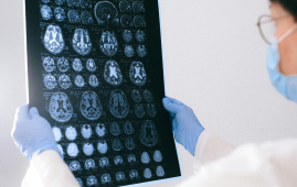
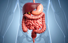
While prior research has suggested that Parkinson’s disease begins in the gut and travels to the brain, the exact mechanism has remained a mystery. A new pre-clinical study led by Duke Health researchers adds to the body of evidence supporting the gut-brain relationship.
The research describes a process in which a protein present in the gut called alpha-synuclein (-synuclein) travels through the nervous system and reaches vulnerable nerves in the brain in a paper published on December 8 in the journal JCI Insight.
“This transport system becomes an issue when alpha-synuclein proteins become corrupted,” said senior author Rodger Liddle, M.D., professor of medicine at Duke University School of Medicine. “If they are corrupted in the gut and are then able to spread to the brain, they could form clumps known as Lewy bodies, which are the hallmark of Parkinson’s disease and other forms of dementia.”
Parkinson’s disease is a degenerative disorder that affects voluntary movement over time. It is predicted that up to ten million people globally are infected.
There is mounting evidence that the stomach plays a role in Parkinson’s disease development. One indicator is that gastrointestinal problems, such as constipation, frequently occur before motor abilities decrease.
Liddle and colleagues concentrated on enteroendocrine cells, which are specialized cells that lining the gut. These cells respond to their surroundings and detect toxicants such as herbicides and pesticides in the intestine; they also contain -synuclein.
The researchers discovered that enteroendocrine cells carry -synuclein from gut mucosal cells to the brainstem via the vagus nerve, the body’s superhighway connecting the stomach and brain.
“We hypothesize that something in the gut is corrupting ⍺-synuclein, causing it to misfold,” he said. We don’t know if this is due to toxicants or another type of exposure.” However, we have shown that pathologically misfolded -synuclein can be transferred from enteroendocrine cells to the brain, where it can accumulate to form Lewy body deposits.
According to Liddle, the study team was able to stop the spread of -synuclein in the rats by cutting the vagus nerve. The discovery lays the groundwork for developing medicines that could either inhibit the transport system or restore the disrupted gut-brain connection.
more recommended stories
 Rheumatoid Arthritis Linked to SPP1hi Macrophages Growth
Key Summary Researchers at Hospital for.
 Resistance Training Reduces Type 2 Diabetes Risk in Adults
Key Takeaways Consistent resistance training was.
 Cardiac Aging Research Identifies PRDM16 Heart Regulator
Key Takeaways Researchers created a single-nucleus.
 Digital Heart Twins Show Promise for AF Ablation Precision
Key Summary Researchers developed patient-specific digital.
 Alzheimer’s Disease Study Questions Glucosamine Safety
Key Summary New research published in.
 WHO: Andes Hantavirus Not a COVID-19-Level Threat
Key Summary Andes hantavirus has drawn.
 Genetic Screening Project Launches for Brazilian Parents
Key Summary Brazil is launching the.
 CAR T-Cell Therapy Offers Hope for Highly Sensitized Patients
Key Summary Researchers at the University.
 Gut Keystone Bacteria: How Diet Shapes Microbiome Health
Key Takeaways Researchers are identifying “gut.
 Chronic Liver Disease in Europe Raises Public Health Alarm
Key Summary A new report in.
Leave a Comment