Image by ArtPhoto_Studio on Freepik
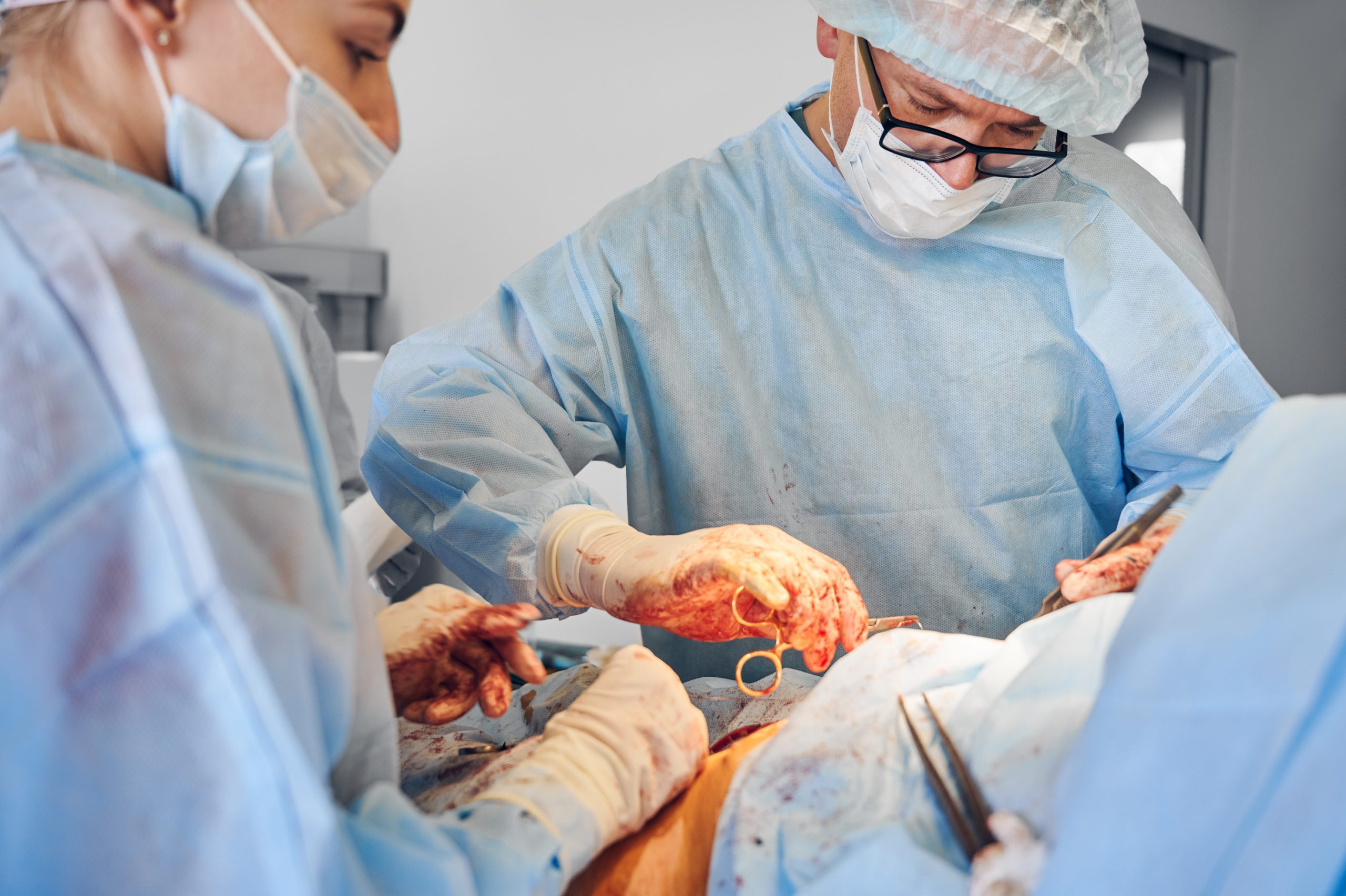
The distinction between cancerous tumors and healthy tissue has now been made for the first time during tumor removal surgery using a novel technology that combines extremely fine-grained, real-time images of inside the body with a particular sort of infrared light.
Engineers from the Wellcome/EPSRC Centre for Interventional and Surgical Sciences (WEISS) at UCL and surgeons at Great Ormond Street Hospital devised the ground-breaking technology tested on mice (GOSH).
The discovery, according to researchers, may have ramifications for the treatment of neuroblastoma, the most frequent solid malignant tumor in children’s bodies outside of brain tumors. Tumor removal surgery is usually required as part of standard care to completely remove malignant cells, which can be hard to spot since they resemble the surrounding healthy tissue.
Researchers from UCL and GOSH utilized a procedure known as “molecular imaging” during surgery, in which chemicals are injected into the bloodstream to serve as imaging probes, for the study, which was published in Cancer Research. Once the probes have been linked to the malignant cells in the body using these chemicals, they begin to fluoresce, which causes the tumor to glow. During preclinical testing in mice, the method successfully exposed a portion of a tumor that had not been surgically excised.
The team then intended to see if employing a “new” type of light, short wave infrared light (SWIR), which has only recently been available to scientists thanks to new technology, could increase the visual clarity of the photographs. To do this, they captured SWIR fluorescence with a specialized high-resolution camera. SWIR is invisible to the naked eye and has a longer wavelength than visible light, allowing it to penetrate deeper into the tissue to generate crisper, more detailed images. During the preclinical studies, this method allowed surgeons to distinguish between malignant tumors and healthy tissue.
Team leader Dr. Stefano Giuliani, Consultant Pediatric Surgeon at Great Ormond Street Hospital and Associate Professor at UCL Great Ormond Street Institute of Child Health, said, “Surgery to remove neuroblastoma requires a delicate balance. Remove too little and the tumor might grow back, but remove too much and the surgeon risks damaging the surrounding blood vessels, nerves, and other healthy organs. This technique effectively lights up the tumor, allowing surgeons to remove it with unprecedented precision. We hope to be able to translate this innovative technology into clinical practice at GOSH as soon as possible to benefit the largest number of children with cancerous tumors.”
A devastating form of juvenile cancer, neuroblastoma accounts for 8–10% of all cases and 15% of all cancer-related fatalities in children. At the time of diagnosis, the cancer has already progressed to about one-third of patients, making treatment more challenging.
Molecular imaging produces precise images of biological processes and can be performed live during surgery, unlike X-rays and magnetic resonance imaging (MRI), which concentrate on organs and bones. As a result, clinical teams don’t have to wait for the results of biopsy or culture when screening for diseases. Real-time picture enhancement is achieved using the SWIR.
Dr. Dale Waterhouse (Wellcome / EPSRC Centre for Interventional & Surgical Sciences (WEISS) at UCL) said, “This work shows that SWIR imaging, a technology first used for material inspection, can enhance the surgeon’s vision beyond the capabilities of the human eye, allowing more precise tumor surgery. It is very exciting to be part of an interdisciplinary team where surgeons and engineers work together, pioneering cutting-edge technologies that promise to improve the treatment of patients at GOSH.”
Dr. Laura Privitera (UCL Great Ormond Street Institute of Child Health) said, “Pediatric surgical oncology faces an ever-increasing need for novel technologies and devices that can help visualize tumors intraoperatively. By using targeted fluorescence-guided surgery, we demonstrate the possibility of safely and specifically delineating tumor margins, allowing its differentiation from surrounding healthy tissue. Fluorescence-guided surgery is a game-changing innovation that will help surgeons to obtain safer and more complete resection. It is exciting to be part of this project, and I look forward to seeing this technology translated into the clinical environment.”
Scientists at GOSH and UCL WEISS are now working to fast-track the technology into the operating theater at GOSH within the next 12 months to benefit children with cancerous tumors.
 Drug Delivery Nanoparticles Studied with New AF4-SANS Tool
Key Points Researchers developed the first.
 Gut-Friendly Diet Linked to Lower CHD Mortality Risk
Key Summary A higher Dietary Index.
 Parkinson’s Disease Risk Linked to Short Sleep, Study Finds
Key Points A large Chinese cohort.
 Cancer Risk And Allergic Diseases Linked in New Study
Key Points A large meta-analysis of.
 Lifestyle Behaviors and Mood Linked in Daily Life Study
Key Points A 70-day diary study.
 Cat Fleas Linked to Murine Typhus Risk in Texas Study
Key Points Researchers detected Rickettsia typhi.
 Pediatric Surgical Safety Reduces Serious Surgical Events
Key Points Pediatric operating room safety.
 England Obesity Prevalence Climbs to 30%, Lancet Study
Key Points A nationwide study of.
 Rheumatoid Arthritis Linked to SPP1hi Macrophages Growth
Key Summary Researchers at Hospital for.
 Resistance Training Reduces Type 2 Diabetes Risk in Adults
Key Takeaways Consistent resistance training was.
Leave a Comment