
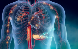
Needle-Thin Brain Implant Enables Precision Neural Drug Delivery
Needle-thin brain implant technology is redefining how researchers study and interact with the brain. A multidisciplinary research team from DTU, the University of Copenhagen, and University College London has introduced a microfluidic Axialtrode (mAxialtrode). This flexible, fiber-based brain implant enables recording of neural signals, optical stimulation, and targeted drug delivery along a single implant shaft.
Published in Advanced Science, this innovation addresses a long-standing challenge in neuroscience: the inability of conventional implants to monitor and modulate multiple brain layers simultaneously. For clinicians and researchers focused on epilepsy, memory, and decision-making disorders, this represents a meaningful shift in experimental precision.
Unlike traditional silicon electrodes that can irritate neural tissue, the mAxialtrode is fabricated from soft, plastic-like polymer fibers using a thermal drawing process similar to ultra-fine fiber pulling. The implant measures less than 0.5 mm in diameter and includes:
Its angled, tapered tip reduces insertion trauma, while its flexibility allows it to move with the brain, an important factor in reducing inflammatory responses seen with rigid implants.
In mouse models, the mAxialtrode demonstrated the ability to:
Notably, animals tolerated the implant well, carrying the lightweight fiber without visible distress. These findings are particularly relevant for epilepsy research, where understanding cross-layer neural signaling is critical.
While further testing and regulatory approvals are required, the researchers are actively pursuing patent protection and evaluating clinical translation pathways. For neurologists, neurosurgeons, and neuroscience researchers, the mAxialtrode introduces a less invasive, multi-functional interface that could shape future strategies in targeted neuromodulation and precision drug delivery.
 SuperAgers and Alzheimer’s Disease Risk Explained
Key Summary SuperAgers maintain exceptional episodic.
 Brain Energy During REM Sleep Shows ATP Paradox
Key Points Researchers identified a unique.
 Intrinsic Cardiac Nervous System Supports Cardiac Stability
Key Points Researchers identified two distinct.
 Multiple Sclerosis Mortality Trends Reveal Care Gaps in the USA
Key Points Multiple sclerosis mortality in.
 Moderate Coffee Intake Associated With Lower CVD Risk
Key Summary Moderate coffee intake (3–5.
 Chronic Xanthan Gum Alters Gut Microbiota and Colon Health
Key Points Chronic xanthan gum consumption.
 Drug Delivery Nanoparticles Studied with New AF4-SANS Tool
Key Points Researchers developed the first.
 Early Menopause Risk Higher in Rural Women: New Study
Key Points Early menopause affects more.
 Parkinson’s Disease Risk Linked to Short Sleep, Study Finds
Key Points A large Chinese cohort.
 Cancer Risk And Allergic Diseases Linked in New Study
Key Points A large meta-analysis of.
Leave a Comment