Title: Dermal features derived from optoacoustic tomograms via machine learning correlate microangiopathy phenotypes with diabetes stage
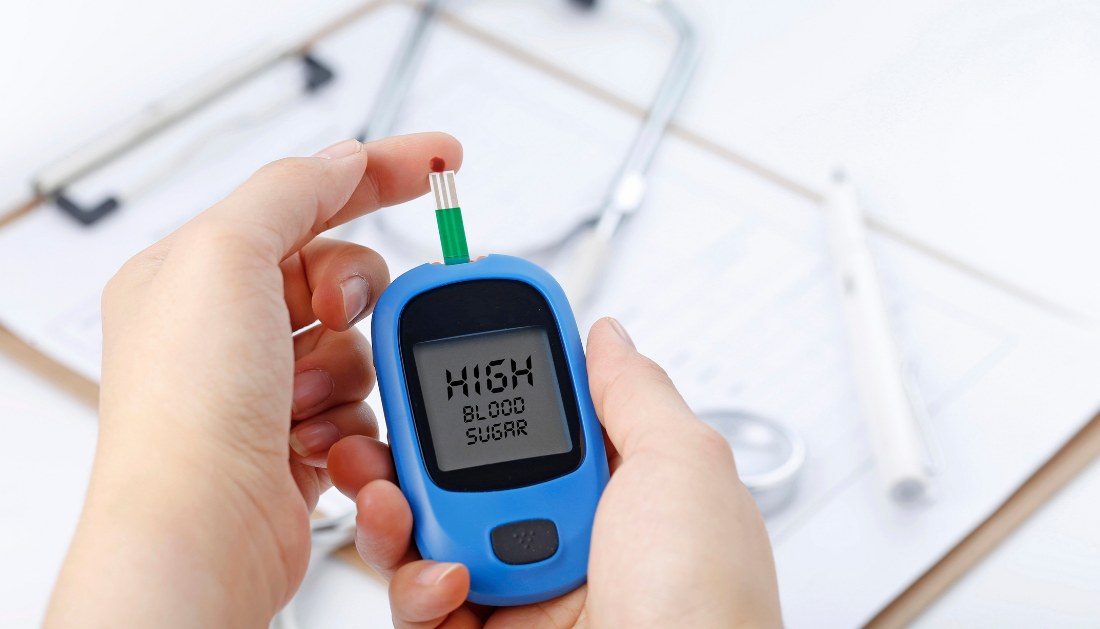
Diabetes is frequently associated with changes in tiny blood arteries. Researchers from the Technical University of Munich (TUM) and Helmholtz Munich have now developed a method for measuring these microvascular alterations in the skin and thereby determining the severity of the condition. They accomplish this by combining artificial intelligence (AI) and cutting-edge high-resolution optoacoustic imaging technologies. The findings have been published in the journal Nature Biomedical Engineering.
Diabetes is frequently associated with changes in tiny blood arteries. Researchers from the Technical University of Munich (TUM) and Helmholtz Munich have now developed a method for measuring these microvascular alterations in the skin and thereby determining the severity of the condition. They accomplish this by combining artificial intelligence (AI) and cutting-edge high-resolution optoacoustic imaging technologies. The findings have been published in the journal Nature Biomedical Engineering.
Light pulses are used to create ultrasound inside tissue in optoacoustic imaging technologies. The resulting ultrasonic waves are then captured by sensors and translated into pictures. The signals are generated by small expansions and contractions of tissue that surround molecules that absorb light strongly. Hemoglobin is one such molecule. Because hemoglobin is concentrated in blood arteries, optoacoustic imaging may create detailed, one-of-a-kind images of vessels that other non-invasive techniques cannot.
Although the fundamental concepts of optoacoustics, or photoacoustics, have been known for over a century, practical applications in medicine are relatively new. Vasilis Ntziachristos is a TUM Professor of Biological Imaging and the Director of the Helmholtz Institute of Biological and Medical Imaging and the Bioengineering Center. He and his colleagues have created several optoacoustic imaging technologies, including RSOM, which stands for raster-scan optoacoustic mesoscopy.
The researchers have now used RSOM to investigate the impact of diabetes on human skin. The researchers used an AI algorithm to identify diabetes characteristics in RSOM pictures of the blood arteries in the legs of 75 diabetics and a control group.
They compiled a list of 32 particularly important modifications based on changes in the appearance of the skin microvasculature. These included characteristics such as the number of branches or the diameter of the vessels.
Biopsies, which are investigations of small portions excised from the skin, have shown that small blood arteries in diabetics’ skin are altered. Biopsies, on the other hand, do not precisely depict live conditions since they can distort blood vessels. They are also intrusive and not appropriate for long-term observations.
RSOM measurements, on the other hand, are not invasive, take less than a minute and do not rely on radiation or contrast agents. “Other optical methods do not achieve the depth or the detail reached by RSOM,” says Angelos Karlas, lead clinician in the study.
Data on several skin depths can be gathered simultaneously with a single RSOM measurement. This allowed the researchers to identify for the first time that diabetes affects arteries differently at different skin layers. For example, while the number of arteries and branches in the dermal layer was reduced in diabetics, it was raised closer to the skin’s surface, in the epidermal layer.
“With RSOM, we can now quantitatively describe the effects of diabetes,” says Vasilis Ntziachristos.”With the emerging ability to make RSOM portable and cost-effective, these findings open up a new way for continuous monitoring of the status of those affected—more than 400 million people worldwide. In the future, with fast and painless examinations, it would take just a few minutes to determine whether therapies are having an effect, even at home environments.”
 Moderate Coffee Intake Associated With Lower CVD Risk
Key Summary Moderate coffee intake (3–5.
 Chronic Xanthan Gum Alters Gut Microbiota and Colon Health
Key Points Chronic xanthan gum consumption.
 Drug Delivery Nanoparticles Studied with New AF4-SANS Tool
Key Points Researchers developed the first.
 Early Menopause Risk Higher in Rural Women: New Study
Key Points Early menopause affects more.
 Cancer Risk And Allergic Diseases Linked in New Study
Key Points A large meta-analysis of.
 Lifestyle Behaviors and Mood Linked in Daily Life Study
Key Points A 70-day diary study.
 Cat Fleas Linked to Murine Typhus Risk in Texas Study
Key Points Researchers detected Rickettsia typhi.
 Pediatric Surgical Safety Reduces Serious Surgical Events
Key Points Pediatric operating room safety.
 Blood Test May Improve Alzheimer’s Disease Prognosis
Key Points A new review suggests.
 Rheumatoid Arthritis Linked to SPP1hi Macrophages Growth
Key Summary Researchers at Hospital for.
Leave a Comment